
8 Ridiculous Radiculopathy Symptoms Mistakes You Don’t Know You’re Making
8 Critical Radiculopathy Symptoms Mistakes Costing You Relief
I’m Dr. Nasser. Radiculopathy symptoms are warning signs that a spinal nerve root is being compressed or irritated. They often include sharp or burning pain that radiates from the neck or back into an arm, chest wall, buttock, or leg, along with numbness, tingling, altered sensation, reflex changes, or muscle weakness such as weak grip or foot drop. These symptoms usually follow a predictable nerve pathway, so the location of the pain or weakness can help identify which nerve root is involved.
Spinal nerves act as communication highways between the central nervous system and the rest of the body. When an exit pathway – known as the neural foramen – narrows due to a disc herniation, bone spurs (osteophytes), or age-related degeneration, the exiting nerve root becomes compressed or chemically irritated. This starts an inflammatory cascade, leading to severe discomfort and possible nerve damage if neglected.
Unfortunately, many people misinterpret what their bodies are telling them. Avoiding the following eight mistakes can help you seek the right care sooner.
Mistake 1: Confusing Sciatica and Radiculopathy Symptoms as the Same Diagnosis
One of the most frequent misconceptions we encounter is the interchangeable use of “sciatica” and “radiculopathy.” While closely related, they are not identical.
Radiculopathy is an umbrella clinical term describing any pinched or inflamed spinal nerve root along the spine. In contrast, sciatica specifically describes compression or irritation affecting the nerve roots that form the sciatic nerve (primarily L4, L5, S1, S2, and S3). Reviewing understanding common sciatica symptoms shows that while all true cases of sciatica represent forms of lumbar radiculopathy, not all radiculopathies are sciatica.
A pinched nerve in your cervical spine (neck) or thoracic spine (mid-back) is a radiculopathy, but it has nothing to do with the sciatic nerve. Assuming every radiating nerve symptom is standard sciatica can lead patients to use the wrong stretches and delay an accurate diagnostic workup.
Mistake 2: Assuming Pinched Nerve Pain Only Happens in the Neck or Lower Back
Because the cervical and lumbar regions bear the most mechanical load and allow the greatest range of motion, they account for the vast majority of spinal complaints. However, assuming nerve roots only get compressed in your lower back or neck leaves a significant blind spot: the thoracic spine.
According to clinical resources from Johns Hopkins Medicine on radiculopathy, the human spine comprises 33 vertebrae divided into distinct segments: cervical, thoracic, lumbar, sacral, and coccygeal. While less common, thoracic radiculopathy accounts for localized and radiating band-like discomfort across the torso.
Focusing only on the extremities means patients often spend months investigating gastrointestinal or cardiovascular issues before someone evaluates their mid-back. Exploring an in-depth guide to lumbar radiculopathy symptoms alongside cervical and thoracic patterns provides a clearer clinical picture of how multi-level nerve involvement presents.
Mistake 3: Overlooking Subtler Cervical Radiculopathy Symptoms Like Grip Weakness
When people think of a pinched nerve in the neck, they typically expect severe, stabbing neck pain. In reality, cervical nerve root compression often presents with subtle distal problems long before the neck itself feels stiff.
C6 and C7 nerve root compressions frequently manifest as:
- Loss of grip strength (dropping coffee mugs, difficulty opening jars)
- Tingling sensation traveling down the outer arm into the thumb, index, or middle finger
- Deep, dull aching around the shoulder blade (scapular boundary) that mimics muscular strain
- Loss of triceps or biceps deep tendon reflexes
Failing to recognize these subtle motor and sensory deficits can allow nerve compression to progress toward functional impairment. Taking time for decoding cervical radiculopathy symptoms ensures that warning signs like grip weakness or localized numbness are caught early.
| Spinal Region | Common Roots Involved | Primary Sensory Patterns | Common Motor / Functional Deficits |
|---|---|---|---|
| Cervical | C5, C6, C7, C8 | Neck, shoulder, lateral arm, thumb, and fingers | Reduced bicep/tricep strength, weak grip, dropping objects |
| Thoracic | T1 through T12 | Band-like torso distribution, rib cage, upper abdomen | Abdominal wall weakness, pain exacerbated by deep breaths |
| Lumbar | L4, L5, S1 | Buttock, anterior thigh, lateral calf, top/sole of foot | Quadricep weakness, foot drop, loss of calf push-off power |
Mistake 4: Dismissing Thoracic Chest Banding as Heart or Gallbladder Issues
Thoracic radiculopathy is relatively rare, but it is one of the most frequently misdiagnosed spinal conditions. Because thoracic spinal nerves travel along the intercostal spaces of the ribs, compression here creates a tight, burning, or band-like sensation wrapping around the rib cage or upper abdomen.
Patients often rush to the emergency room fearing a heart attack, pulmonary embolism, or acute gallbladder attack. While ruling out life-threatening cardiac and visceral conditions is necessary, once internal organ pathologies are cleared, the spine should be examined.
Thoracic nerve roots can become irritated by degenerative changes, disc herniations, spinal trauma, or microvascular complications such as diabetic nerve root ischemia.
Mistake 5: Ignoring Red-Flag Warning Signs Like Saddle Anesthesia and Bladder Changes
While most cases of radiculopathy resolve with conservative, non-surgical management, certain “red flag” presentations represent medical emergencies requiring immediate surgical decompression. Ignoring these symptoms can lead to permanent neurological damage, paralysis, or lifelong incontinence.
You must seek urgent medical evaluation if you experience:
- Saddle Anesthesia: Sudden numbness or loss of sensation in the groin, buttocks, inner thighs, or perineum.
- Bowel or Bladder Dysfunction: Sudden urinary retention, overflow incontinence, or loss of bowel sphincter control.
- Progressive Motor Deficits: Rapidly worsening motor loss, such as an inability to lift the front part of the foot – learn more about managing L5 nerve root foot drop to prevent tripping hazards.
- Bilateral Neurological Symptoms: Severe weakness or sensory loss progressing across both legs simultaneously, which may indicate cauda equina syndrome or severe spinal cord compression (myelopathy).
Mistake 6: Relying Strictly on MRIs Without Proper Provocative Physical Testing
Magnetic Resonance Imaging (MRI) is an exceptional diagnostic tool, but treating an MRI image rather than the patient is a major mistake. As we age, spinal discs naturally dehydrate, lose height, and bulge. Many adults over 40 have asymptomatic disc herniations or bone spurs on their scans that cause zero nerve inflammation or pain.
According to medical research on evidence-based diagnostic criteria for radiculopathy published via the NCBI Bookshelf, imaging findings must always correlate directly with clinical physical examination maneuvers.
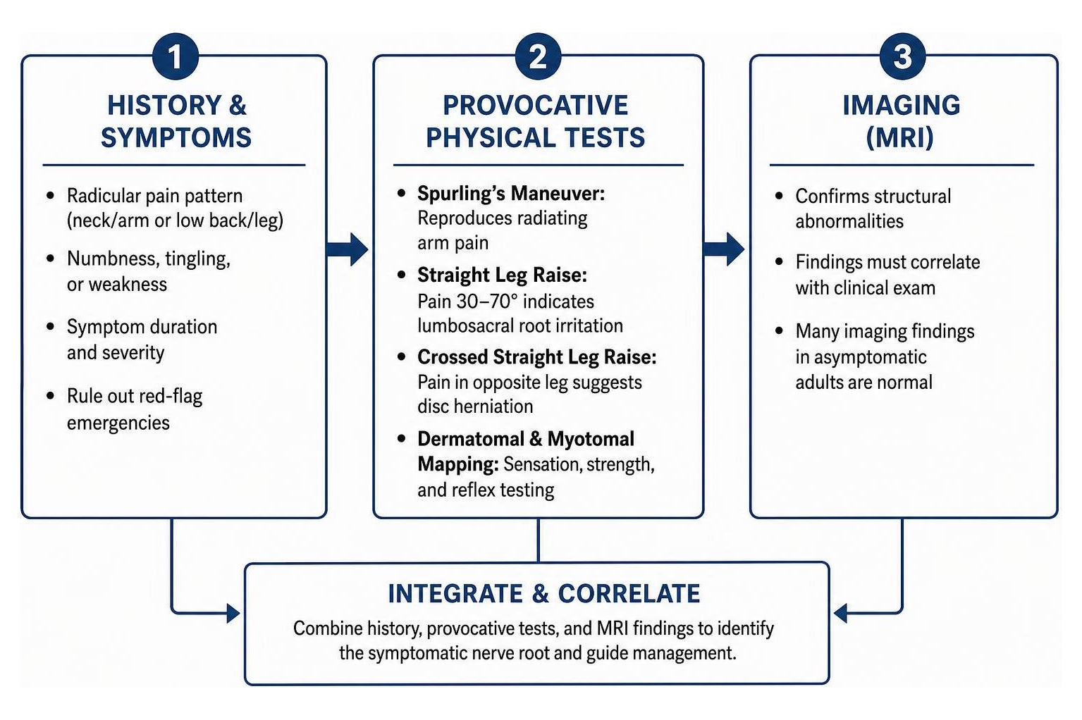
A comprehensive evaluation requires provocative tests:
- Spurling’s Maneuver: Gently extending, rotating, and applying axial compression to the cervical spine to see if it reproduces radiating arm pain.
- Straight Leg Raise (Lasegue’s Sign): Raising the extended leg of a supine patient between 30 and 70 degrees to traction the lumbosacral roots.
- Crossed Straight Leg Raise: Raising the unaffected leg; if pain radiates down the opposite symptomatic leg, it is highly specific for a disc herniation.
- Dermatomal and Myotomal Mapping: Testing pinprick sensation, motor resistance, and reflex symmetry to pinpoint the compressed nerve root.
Mistake 7: Believing Surgery Is the Immediate and Only Lasting Fix
Hearing that a spinal nerve is “pinched” leads many patients to assume their spine requires immediate surgical restructuring. In truth, surgery is rarely the first-line answer.
Clinical studies demonstrate that 75% to 90% of patients with acute radiculopathy recover using conservative treatment plans (Berry et al., 2019). The body possesses an innate ability to heal: herniated disc fragments often undergo natural enzymatic breakdown, dehydration, and macrophage-driven reabsorption over several months.
Starting with interventional pain management for lumbar radiculopathy – such as targeted fluoroscopic epidural steroid injections, selective nerve root blocks, and structured physical therapy – helps calm inflamed nerve roots while preserving natural spinal biomechanics.
Mistake 8: Masking Nerve Root Inflammation with NSAIDs Long-Term
Over-the-counter nonsteroidal anti-inflammatory drugs (NSAIDs) such as ibuprofen, naproxen, and meloxicam can provide temporary symptom relief during acute flare-ups. However, relying on them as a long-term plan is risky.
Using NSAIDs consecutively beyond 10 to 14 days without physician supervision significantly elevates the risk of peptic ulcer disease, gastrointestinal bleeding, renal stress, and cardiovascular complications. Furthermore, NSAIDs only dampen surrounding tissue inflammation without addressing deep neuropathic pain signals or structural neuroforaminal narrowing.
A comprehensive plan may include a short oral corticosteroid taper, membrane-stabilizing neuropathic modulators (like gabapentin or pregabalin), and targeted cervical radiculitis treatment to resolve inflammation safely at the source.
How Spinal Specialists Accurately Diagnose Pinched Nerve Roots
Accurately identifying the exact source of radiating pain requires a structured clinical approach. At Pain Arizona, our double board-certified physicians combine clinical examinations with advanced diagnostic technology.
- Detailed Clinical History & Symptom Chronology: We review when the pain began, what maneuvers worsen or relieve it (such as the shoulder abduction relief test for neck pain), and map the distribution of paresthesias.
- Neurological Examination: We perform sensory testing across specific dermatomes, test muscle motor strength across corresponding myotomes, and evaluate deep tendon reflexes (biceps, triceps, brachioradialis, patellar, and Achilles).
- Advanced Diagnostic Imaging: Non-contrast MRI remains the gold standard for visualizing soft-tissue disc herniations, the spinal cord, and exit foramina. For patients who cannot undergo MRI due to pacemakers or metal hardware, CT myelography provides detailed cross-sectional views of bone and nerve structures.
- Electrodiagnostic Studies (EMG/NCS): Electromyography and Nerve Conduction Studies evaluate the electrical integrity of muscles and peripheral nerves. As highlighted in clinical resources from Merck Manuals on nerve root disorders, electrodiagnostics help differentiate true root-level compression (radiculopathy) from distal peripheral nerve entrapments (such as carpal tunnel syndrome) or systemic diabetic peripheral neuropathy.
Evidence-Based Treatment Pathways for Lasting Decompression
Once a precise diagnosis is established, treatment focuses on reducing inflammation, relieving mechanical compression, and restoring pain-free functional movement.
1. Interventional Pain Management Procedures
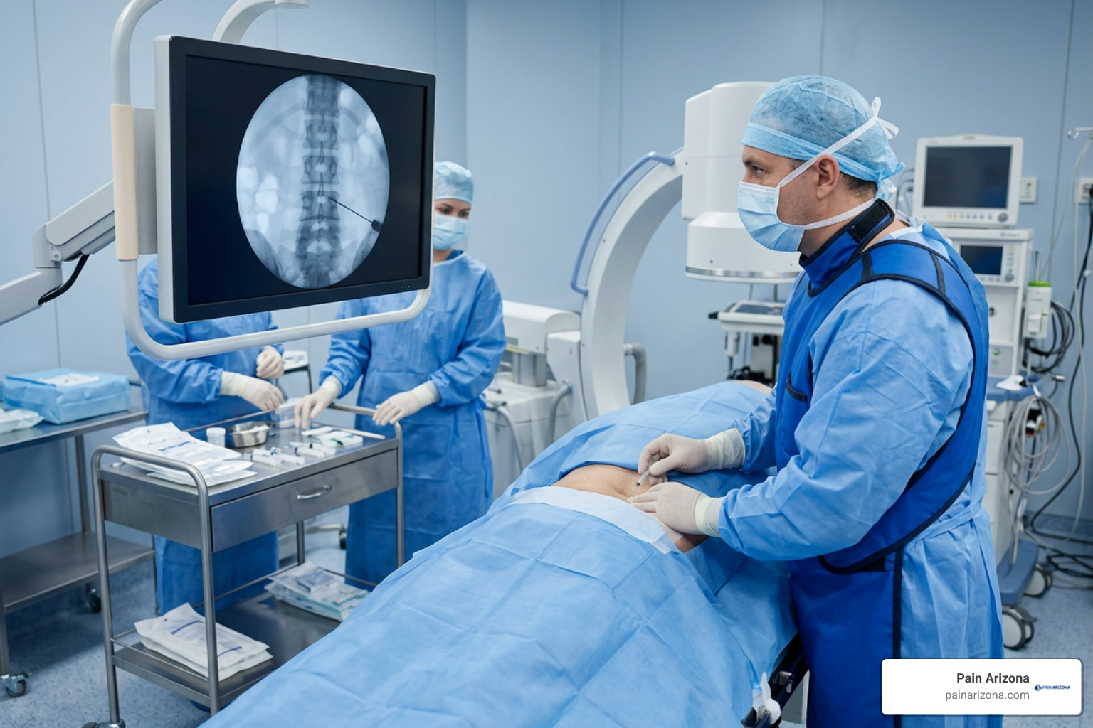
When oral medications and rest do not provide sufficient relief, image-guided interventional procedures deliver targeted anti-inflammatory agents directly to the affected nerve root:
- Transforaminal Epidural Steroid Injections (TFESI): Performed under live fluoroscopic (X-ray) guidance, this procedure places a concentrated corticosteroid and local anesthetic solution directly into the neuroforaminal space where the nerve is compressed.
- Selective Nerve Root Blocks (SNRB): Both therapeutic and diagnostic, SNRBs help isolate single nerve roots to confirm pain generators and alleviate radicular pain.
- Radiofrequency Ablation (RFA): If facet joint arthritis or spondylosis contributes to spinal pain and functional restriction, radiofrequency neurotomy can disrupt pain signals from the medial branch nerves.
2. Structured Physical Rehabilitation
Exercise therapy must be tailored to the patient’s specific directional preference and spinal pathology. Following structured physical therapy protocols for cervical radiculopathy ensures spinal nerves are gently mobilized without aggravating inflammation.
Core rehabilitation components include:
- McKenzie Extension and Retraction Exercises: Movements designed to centralize radiating pain back toward the spine.
- Neural Flossing / Nerve Gliding: Gentle, dynamic stretches that facilitate smooth motion of spinal nerves through tight fibrous tunnels.
- Cervical Deep Neck Flexor Conditioning: Strengthening longus colli and longus capitis muscles to reduce cervical lordotic strain.
- Core and Lumbar Stabilization: Activating the transversus abdominis, multifidus, and pelvic floor muscles to reduce excessive mechanical loading on the lumbar discs.
- Postural Ergonomics: Adjusting computer workstation heights, lumbar supports, and driving postures to minimize sustained neuroforaminal pinching.
3. Surgical Decompression (When Indicated)
If a patient experiences severe, progressive motor deficits (such as worsening foot drop or hand muscle atrophy), red-flag cauda equina symptoms, or intractable pain that fails to improve after 6 to 8 weeks of comprehensive conservative care, surgical options like a minimally invasive microdiscectomy, foraminotomy, or anterior cervical discectomy and fusion (ACDF) may be discussed (Alexander et al., 2024).
Frequently Asked Questions about Radiculopathy Symptoms
Does radiculopathy ever go away on its own?
Yes. Many mild to moderate cases of acute radiculopathy resolve within 6 to 12 weeks with conservative care. Natural disc reabsorption, local biochemical resolution of inflammatory cytokines, and posture correction help decompress the nerve root over time.
What is the primary difference between radiculopathy, neuropathy, and myelopathy?
These conditions affect different parts of the nervous system:
- Radiculopathy: Irritation or pinching of a spinal nerve root where it exits the spine, creating asymmetric symptoms along a specific dermatomal path. Deep tendon reflexes in the affected limb are typically diminished or absent (hyporeflexia).
- Neuropathy (Peripheral): Damage to peripheral nerves away from the spine (common in diabetes), usually causing symmetric, “glove-and-stocking” numbness, burning, and tingling in both hands or feet.
- Myelopathy: Compression of the spinal cord itself inside the spinal canal. This central nervous system condition typically causes balance issues, clumsiness, spasticity, hyperreflexia (exaggerated reflexes), and requires prompt surgical evaluation.
How long does nerve root recovery typically take after treatment?
Nerves heal gradually, typically regenerating at an approximate rate of 1 millimeter per day. Following an epidural steroid injection or the start of structured physical therapy, pain relief can begin within a few days to two weeks. However, recovering lost sensation or resolving deep muscle weakness can take several months depending on the severity and duration of the initial compression.
Finding Lasting Relief with Pain Arizona
Living with radiating nerve pain, tingling, or weakness can make daily activities exhausting. At Pain Arizona, our double board-certified physicians specialize in interventional pain medicine and physical medicine and rehabilitation. We focus on diagnosing the precise structural cause of your pain and building a comprehensive, non-surgical relief plan tailored to your goals.
Whether you are managing chronic lower back issues, exploring evaluating advanced bulging disc treatments, or dealing with new numbness radiating down your arm, our team is here to help you get back to the activities you enjoy. We have convenient clinic locations across the Greater Phoenix Area, including Phoenix, Scottsdale, Mesa, Gilbert, and Glendale.
We accept most insurance plans. Check with our office manager to confirm whether your specific plan is accepted, and schedule your evaluation today.
References
Berry JA, Elia C, Saini HS, Miulli DE. “A Review of Lumbar Radiculopathy, Diagnosis, and Treatment..” Cureus, 2019. PMCID PMC6858271. Christopher E. Alexander, Luke J. Weisbrod, Matthew A. Varacallo. “Lumbosacral Radiculopathy.” StatPearls Publishing, 2024. NBK430837.