
In-Depth Guide to Lumbar Radiculopathy Symptoms
Understanding Lumbar Radiculopathy and Its Causes
I’m Dr. Nasser. To understand lumbar radiculopathy, we first have to look at the architecture of your lower back. Your lumbar spine consists of five large vertebrae (labeled L1 through L5). Between these bones sit intervertebral discs, which act as shock absorbers.
Nerve roots exit the spinal canal through small openings called “foramina.” When anything narrows that opening or encroaches on the space where the nerve sits, the nerve becomes compressed. This is what we mean when we say a nerve is “pinched.”
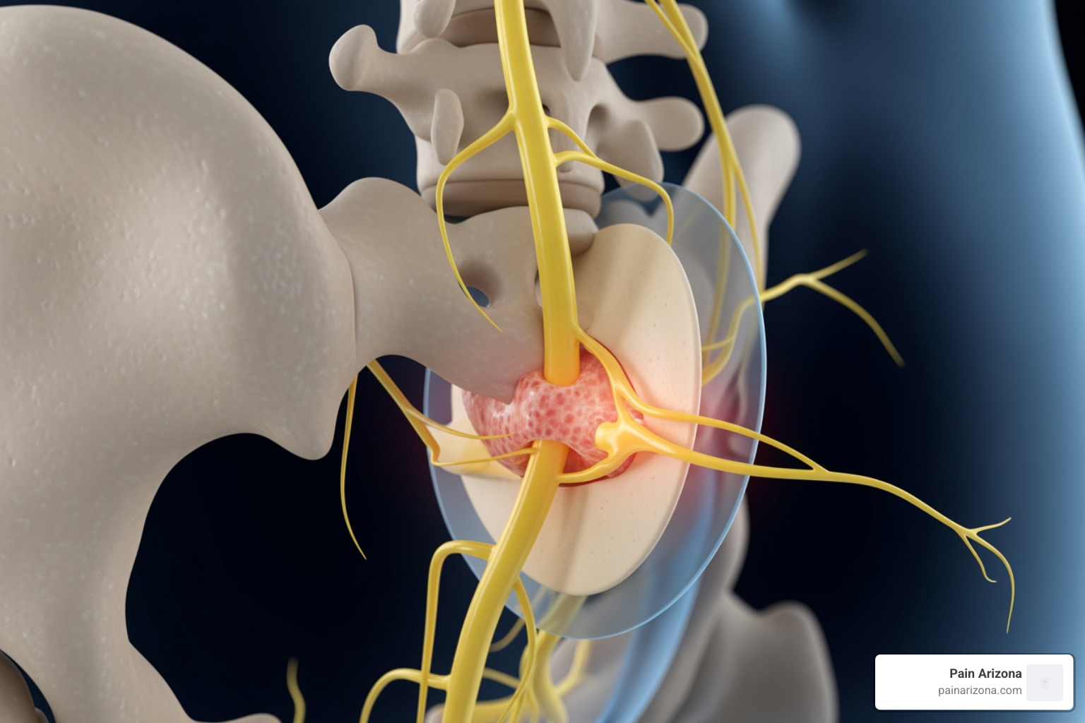
While many factors can lead to this compression, the most frequent culprits we see at Pain Arizona include:
- Disc Herniation: This is the most common cause. The soft, jelly-like center of a disc pushes through a tear in the tougher exterior, directly pressing on the nearby nerve root.
- Foraminal Stenosis: This is a narrowing of the holes (foramina) where the nerves exit. It is often a result of aging and “wear and tear.”
- Degenerative Spondyloarthropathies: As we age, the joints in our spine (facet joints) can develop arthritis. This leads to bone spurs (osteophytes) that poke into the nerve space.
- Ligamentum Flavum Thickening: Ligaments inside the spinal canal can thicken over time, reducing the available room for nerves.
- Spondylolisthesis: This occurs when one vertebra slides forward over the one below it, which can stretch or pinch the nerve roots.
According to research from StatPearls, most disc herniations occur posterolaterally because the posterior longitudinal ligament is strongest in the midline, forcing the disc material toward the nerve roots. For more on the basics, you can read our guide on Understanding Lumbar Radiculopathy.
Differentiating Lumbar Radiculopathy from Sciatica and Neuropathy
Patients often come into our Phoenix and Gilbert offices using the term “sciatica.” While people use them interchangeably, they aren’t exactly the same thing.
- Sciatica: This is a general term for pain that travels along the sciatic nerve (the largest nerve in the body). Most sciatica is actually caused by lumbar radiculopathy at the L4, L5, or S1 levels.
- Peripheral Neuropathy: This is damage to the nerves after they have left the spine area, often caused by diabetes or toxins. It usually feels like a “stocking-glove” pattern (numbness in both feet or both hands) rather than a single stripe of pain down one leg.
- Cervical and Thoracic Radiculopathy: These are “pinched nerves” in the neck or middle back, respectively. Thoracic radiculopathy is actually the rarest form.
- Myelopathy: This is a more serious condition where the actual spinal cord is compressed. This can cause balance issues and coordination problems, whereas radiculopathy affects the individual nerve roots.
| Feature | Radiculopathy | Neuropathy | Myelopathy |
|---|---|---|---|
| Primary Location | Nerve root (Spine) | Peripheral nerves (Limbs) | Spinal Cord |
| Pain Pattern | Follows a dermatome (stripe) | Symmetrical (socks/gloves) | Generalized weakness/balance |
| Reflexes | Decreased or absent | Often normal/decreased | Increased (hyperreflexia) |
| Cause | Disc herniation, stenosis | Diabetes, alcohol, chemo | Severe stenosis, trauma |
Identifying Symptoms, Red Flags, and Diagnostic Pathways
The hallmark of lumbar radiculopathy is “radicular pain.” This isn’t just a dull ache; patients often describe it as an electric shock, a searing burn, or a sharp stabbing sensation.
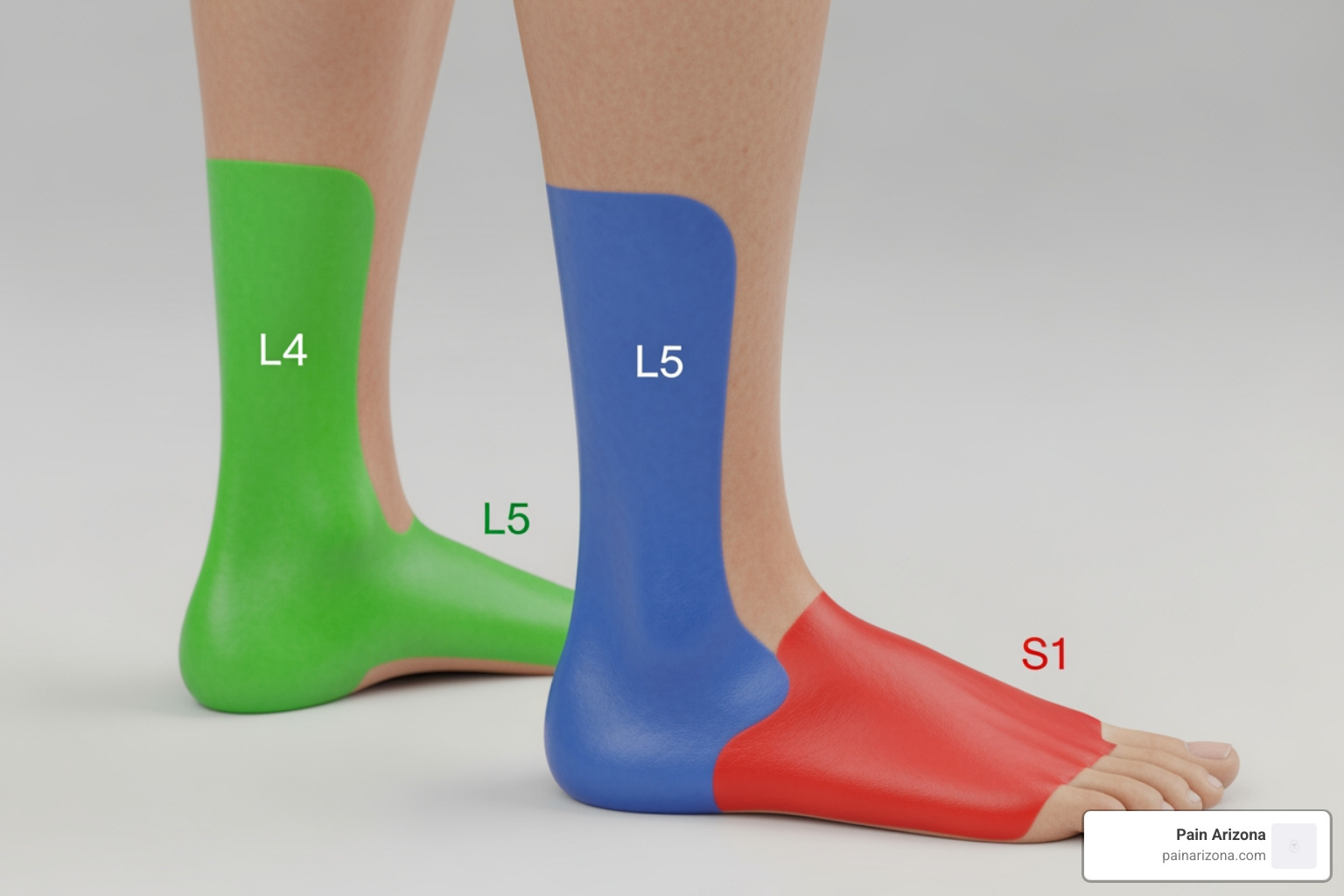
Because each nerve root supplies a specific area of skin (a dermatome) and specific muscles (a myotome), we can often tell exactly which nerve is pinched just by where you feel the symptoms.
- L4 Nerve Root: Pain usually travels across the front of the thigh and down to the inner side of the lower leg. You might have trouble straightening your knee.
- L5 Nerve Root: This is very common. Pain travels down the side of the leg to the top of the foot and the big toe. A classic sign is foot drop, where you struggle to lift your toes toward your shin.
- S1 Nerve Root: Pain travels down the back of the calf to the outer edge of the foot and the pinky toe. You might find it hard to push off your foot while walking.
Beyond pain, you may experience paresthesia (pins and needles), numbness, or a loss of the “jerk” reflex in your knee or ankle. For a deeper dive into these sensations, see Everything You Need to Know About Radiculopathy Treatment.
Recognizing Red Flags and Cauda Equina Syndrome
While most cases of lumbar radiculopathy are not emergencies, there are “red flags” that require immediate medical attention. If you experience any of the following, please head to the nearest emergency room in the Greater Phoenix Area:
- Saddle Anesthesia: Numbness in the areas that would touch a horse saddle (groin, buttocks, inner thighs).
- Bladder or Bowel Incontinence: Inability to hold your urine or stool, or inability to go when you need to.
- Sudden Paresis: Rapidly worsening weakness in one or both legs (e.g., your leg suddenly “giving out”).
- Severe Night Pain: Pain that is worse when lying flat and prevents sleep entirely.
These symptoms can indicate Cauda Equina Syndrome, a rare but surgical emergency where the bundle of nerve roots at the end of the spinal cord is severely compressed. You can find more technical details on these emergencies in the Merck Manual.
Clinical Evaluation and Diagnostic Testing
When you visit us at Pain Arizona, our diagnostic process begins with a physical exam. We use “neural tension tests” to see if moving your leg provokes the nerve.
- Straight-Leg Raise (Lasegue’s Sign): While you lie flat, we lift your straight leg. If you feel shooting pain between 30 and 70 degrees, it’s a strong indicator of a disc herniation.
- Slump Test: While seated, you slump forward and straighten your leg. This is a more sensitive test for nerve tension.
Imaging and Tests:
- MRI: This is the “gold standard.” It allows us to see the discs, nerves, and ligaments in high definition. However, we always correlate the MRI with your symptoms, as many healthy people have disc bulges on an MRI that don’t cause any pain.
- CT Myelogram: Used if you cannot have an MRI (for example, if you have a pacemaker).
- EMG/NCS (Electrodiagnostic Studies): These “nerve tests” check the electrical activity of your muscles and nerves. They are helpful if the diagnosis is unclear or if we need to see how much damage the nerve has sustained. Note: These tests often don’t show abnormalities until you’ve had symptoms for at least 2–3 weeks.
If you are in the middle of a sudden flare-up, check out our Acute Lumbosacral Radiculopathy Guide for immediate steps.
Evidence-Based Treatment and Surgical Indications
The good news? Roughly 90% of people with a disc herniation see significant improvement within 3 months without surgery. We almost always start with conservative management.

Conservative Options Include:
- Activity Modification: We don’t recommend total bed rest (that can actually make things stiffer). Instead, we suggest “relative rest”—staying active but avoiding heavy lifting or twisting.
- Medications: NSAIDs (like ibuprofen) help with inflammation. In some cases, we may use oral corticosteroids or neuropathic medications like gabapentin to quiet the irritated nerve.
- Epidural Steroid Injections (ESIs): If medications and therapy aren’t enough, we can perform a transforaminal epidural injection. This delivers powerful anti-inflammatory medication directly to the “pinch point.” As noted in PMC research, these injections can be both diagnostic (confirming the pain source) and therapeutic.
Physical Therapy and McKenzie Exercises for Lumbar Radiculopathy
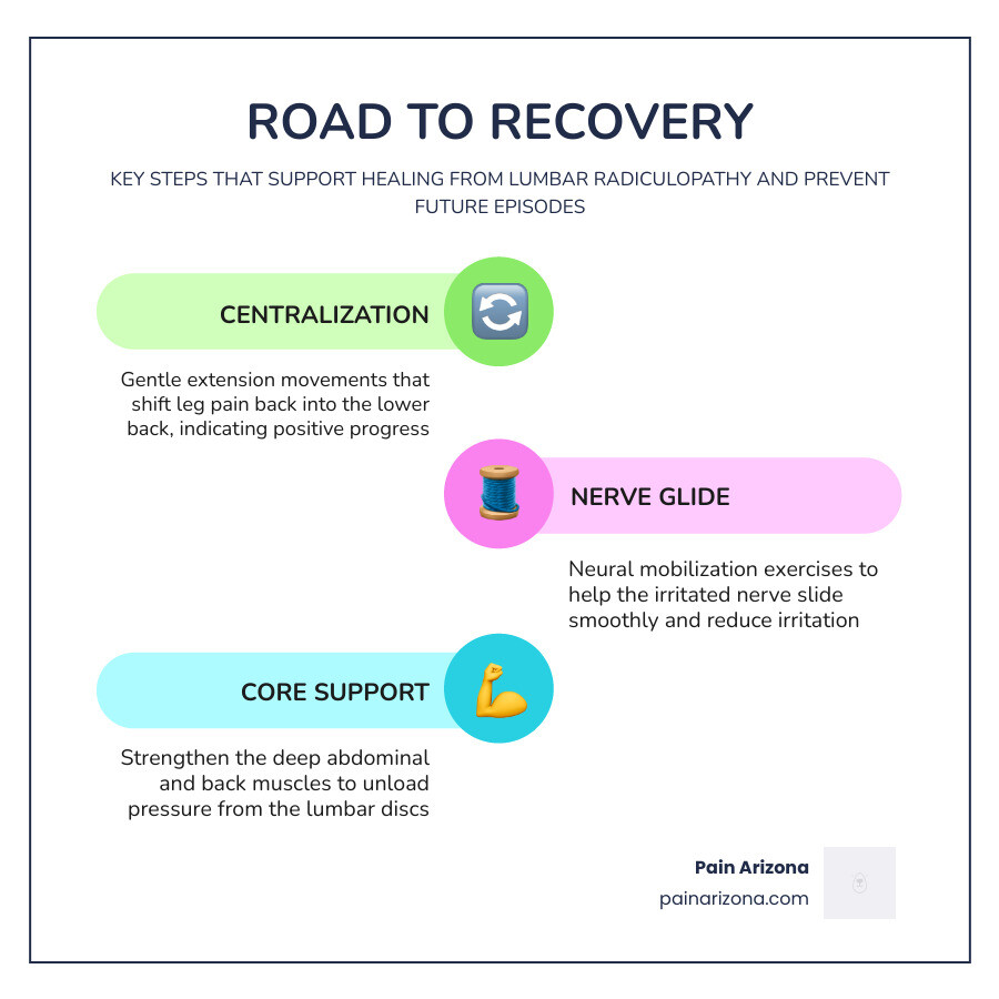
Physical therapy is a cornerstone of recovery. We often focus on “directional preference”—finding a specific movement that makes the pain move out of your leg and back into your lower back. This process is called centralization, and it’s a very positive sign for recovery.
- McKenzie Exercises: Often involve gentle backward bending (extensions) to help “tuck” the disc material back away from the nerve.
- Neural Mobilization: Gentle “flossing” movements that help the nerve glide more smoothly through the spinal canal.
- Core Strengthening: Building the “natural corset” of muscles around your spine to take the pressure off your discs.
Learn more about these techniques in our article on Effective Physical Therapy for Lumbar Radiculopathy.
When to Consider Surgical Intervention
We generally reserve surgery for patients who have failed 6–12 weeks of conservative care or those with progressive neurological deficits (like a worsening foot drop).
The most common procedure is a microdiscectomy, where a surgeon uses a microscope to remove only the small piece of disc that is pressing on the nerve. The landmark SPORT trial showed that while surgery often provides faster relief, both surgical and non-surgical patients tend to have similar outcomes after two years.
Surgery is most beneficial for patients with “sequestered fragments” (a piece of disc that has broken off) or those with severe, intractable leg pain that prevents any quality of life. For a full breakdown of surgical vs. non-surgical paths, see our L5 Radiculopathy Treatment Complete Guide.
Prognosis and Long-Term Prevention
The prognosis for lumbar radiculopathy is generally excellent. About 50% of cases resolve within 1–2 weeks, and the vast majority are better within 3 months. However, low back pain is a leading cause of workplace absenteeism, so long-term management is key to preventing a recurrence.
How to stay pain-free:
- Weight Management: Every extra pound puts more pressure on your lumbar discs.
- Posture and Ergonomics: If you work in an office in Mesa or Scottsdale, ensure your chair supports your lumbar curve. Break up long periods of sitting every 30 minutes.
- Smart Lifting: Never bend at the waist to pick up heavy objects. Use your legs and keep the object close to your body.
- Smoking Cessation: Smoking reduces blood flow to the spinal discs, making them more prone to drying out and tearing.
- Regular Exercise: Walking, swimming, and yoga are fantastic for keeping the spine flexible and supported.
Frequently Asked Questions about Lumbar Radiculopathy
How long does it take for lumbar radiculopathy to heal?
Most people feel significantly better within 6 to 12 weeks. While the “lightning” pain often fades first, some tingling or mild numbness can linger for a few months as the nerve heals.
Can lumbar radiculopathy go away on its own?
Yes. The body has a natural ability to “resorb” herniated disc material. Over time, the immune system recognizes the disc fragment as foreign and breaks it down, which relieves the pressure on the nerve.
What is the difference between a bulging disc and radiculopathy?
A bulging disc is a structural finding (the “what”), while radiculopathy is the clinical syndrome of symptoms (the “result”). You can have a bulging disc that doesn’t touch a nerve, in which case you wouldn’t have radiculopathy.
Conclusion
Lumbar radiculopathy can be a frightening and painful experience, but it is a condition that we successfully manage every day at Pain Arizona. Whether you are dealing with a new “tweak” in your back or chronic radiating leg pain, our goal is to get you back to your life in the Phoenix Area without unnecessary surgery.
Our team of double board-certified physicians specializes in evidence-based care, from advanced physical therapy protocols to minimally invasive injections. We accept most insurance plans; please check with our office manager to see if your plan is accepted.
If you’re tired of the “electric” pain holding you back, don’t wait for the nerve damage to become permanent. Request an Appointment at one of our locations in Phoenix, Gilbert, or across the Valley today.