
Acute Lumbosacral Radiculopathy: Conquer Pain in 4-6 Wks
Understanding Acute Lumbosacral Radiculopathy: What You Need to Know
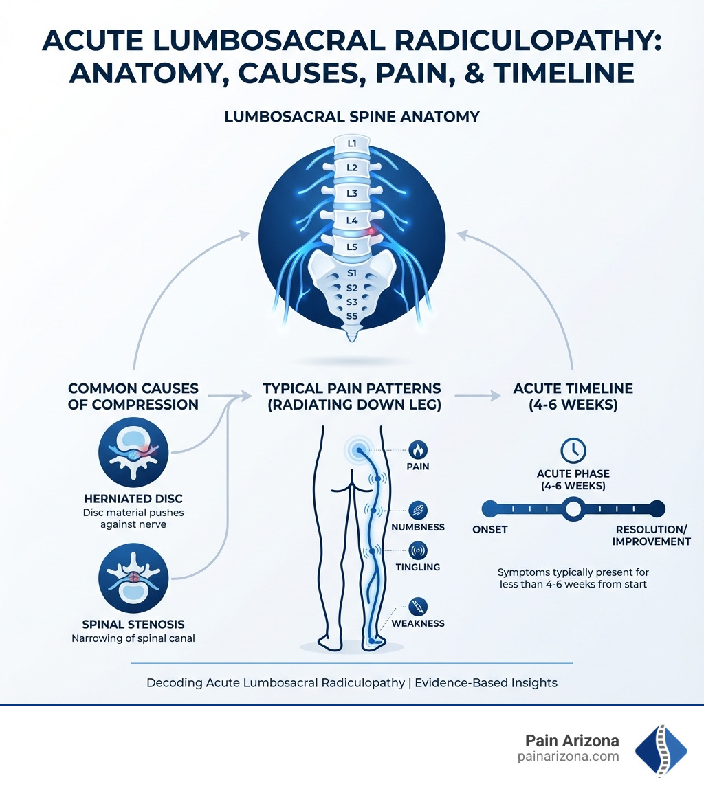
Acute lumbosacral radiculopathy is a condition where one or more nerve roots in your lower back become compressed or irritated, causing pain that radiates down your leg. Here’s what defines this condition:
- What it is: Nerve root compression or irritation in the lower (lumbar) spine
- Main symptoms: Sharp or burning leg pain, numbness, tingling, and weakness
- “Acute” timeline: Symptoms present for less than 4-6 weeks from onset
- How common: Affects 3-5% of the adult population
- Key difference: Unlike general back pain, radiculopathy involves nerve damage causing symptoms that travel down the leg
Most people experience this as sudden, shooting pain that starts in the lower back and travels down one leg. You might also notice numbness in your foot, weakness when trying to lift your toes, or a “pins and needles” sensation. The good news? 70-80% of people improve within 4-6 weeks with proper management.
The pain happens when something—usually a herniated disc, bone spur, or narrowing of the spinal canal—presses on a nerve root as it exits your spine. Think of it like a kink in a garden hose. The nerve can’t send signals properly, so you feel pain, numbness, or weakness in the areas that nerve controls.
I’m Dr. Nasser, Chief Medical Officer at Pain Arizona, and I’ve spent over 15 years helping patients find relief from acute lumbosacral radiculopathy through evidence-based treatments ranging from conservative care to advanced minimally invasive procedures. My goal is to help you understand what’s causing your symptoms so we can create a personalized plan to get you back to the activities you love.
Understanding the Causes and Risk Factors of Acute Lumbosacral Radiculopathy
When we talk about acute lumbosacral radiculopathy, we’re primarily referring to a situation where a nerve root in your lower back is being compressed or irritated. This compression can lead to inflammation, which in turn causes the characteristic pain, numbness, and weakness that define the condition. Imagine the delicate nerve root being squeezed or rubbed by surrounding structures – that’s essentially what’s happening.
Primary Causes
Several structural issues can put pressure on these vital nerve roots:
- Herniated Discs: This is arguably the most common culprit behind acute lumbosacral radiculopathy. Our spinal discs act as cushions between vertebrae. When the soft inner material of a disc pushes through its tougher outer layer, it can press directly on a nearby nerve root. You can learn more about this common condition on our page about Herniated Discs.

- Spinal Stenosis: This condition involves the narrowing of the spinal canal, which houses the spinal cord and nerve roots. This narrowing can be caused by various factors, including thickened ligaments, enlarged facet joints, or bone spurs. When the space becomes too constricted, it can pinch the nerve roots, leading to symptoms. We offer comprehensive information on Spinal Stenosis and its management.
- Degenerative Disc Disease: As we age, our spinal discs naturally lose water content and become less flexible. This “wear and tear” can lead to disc height loss, bulging, or even herniation, all of which can contribute to nerve compression. Understanding Degenerative Disc Disease: Aging and Back Health is key to grasping this cause.
- Bone Spurs (Osteophytes): These bony growths can develop on the vertebrae as a response to disc degeneration or arthritis. While often harmless, they can sometimes grow into the spinal canal or foramina (the openings where nerve roots exit), impinging on a nerve.
- Spondylolisthesis: This occurs when one vertebra slips forward over the one below it. This misalignment can reduce the space available for nerve roots, leading to compression and symptoms of radiculopathy. Our page on Spondylolisthesis provides more details.
- Trauma or Work Injuries: Sudden impacts, falls, or repetitive strain injuries can damage spinal structures, leading to disc herniations or other issues that compress nerve roots. This is particularly relevant for those who experience Work Injuries in physically demanding jobs.
Associated Risk Factors
While some causes are structural, certain factors can increase your likelihood of developing acute lumbosacral radiculopathy:
- Age: This condition is often age-related. According to the National Institutes of Health, men tend to notice symptoms in their 40s and women in their 50s. The overall prevalence is higher in people over 50.
- Genetics: If your family has a history of spinal deformities or weaknesses, you might be more predisposed.
- Occupation: Jobs that involve frequent heavy lifting, especially with twisting motions, prolonged sitting (like truck driving), or heavy industry work can put extra strain on the lower back, increasing risk.
- Sedentary Lifestyle: Lack of physical activity can weaken core muscles that support the spine, making it more vulnerable to injury.
- Obesity: Excess body weight places additional stress on the lower back, contributing to disc degeneration and compression.
- Smoking: Smoking can impair blood flow to spinal discs, accelerating their degeneration and making them more prone to herniation.
- Other Factors: The American Academy of Physical Medicine and Rehabilitation highlights other risk factors such as back trauma, taller height, multiple pregnancies, a history of back pain, and even chronic cough. Maintaining a healthy weight and staying fit are crucial preventative measures.
Diagnosis: Identifying Symptoms and Getting the Right Evaluation
When you experience symptoms suggestive of acute lumbosacral radiculopathy, a thorough clinical evaluation is our first step. This involves taking a detailed patient history and conducting a comprehensive physical examination. We want to understand not just your pain, but how it impacts your daily life in Phoenix, Mesa, or Glendale.
Typical Symptoms of Acute Lumbosacral Radiculopathy
The hallmark of acute lumbosacral radiculopathy is pain that radiates from the lower back into the leg. This isn’t just a dull ache; it’s often described as:
- Radiating Pain: Sharp, burning, lancinating, or stabbing sensations that travel down the leg. This pain can follow specific pathways.
- Dermatomal Patterns: The pain, numbness, or tingling typically follows a specific dermatome, which is an area of skin supplied by a single nerve root. For example:
- L4 radiculopathy: May cause pain and numbness in the front of the thigh and inner calf, potentially affecting the knee jerk reflex.
- L5 radiculopathy: Often results in pain radiating down the outside of the thigh and leg, into the top of the foot and big toe. This is the single most common lumbar radiculopathy.
- S1 radiculopathy: Can cause pain down the back of the thigh and calf, to the sole of the foot and little toe, often affecting the ankle jerk reflex.
- Numbness and Tingling: You might feel a “pins and needles” sensation or a complete loss of feeling in the affected dermatome.
- Muscle Weakness: Depending on the nerve root involved, you might experience weakness in specific muscle groups. For instance, L5 compression can lead to a “foot drop,” making it difficult to lift the front of your foot. While muscle strength can be preserved in milder cases, severe compression can significantly impact it.
- Loss of Reflexes: Specific nerve roots control certain reflexes. A diminished or absent knee jerk (L3-L4) or ankle jerk (L5-S1) reflex can indicate nerve root involvement.
- Pain Worsened by Coughing or Sneezing: Activities that increase pressure in your abdomen, like coughing, sneezing, or even straining during a bowel movement, can temporarily intensify the pain because they increase pressure around the nerve roots.
Diagnostic Process and Tools
Our diagnostic approach is designed to accurately pinpoint the source of your pain:
- Physical Exam Tests: We perform specific maneuvers to assess nerve root irritation:
- Straight Leg Raise (Lasègue test): Lying on your back, we gently raise your affected leg. If this reproduces your leg pain, it suggests nerve root compression.
- Crossed Straight Leg Raise: Raising the unaffected leg causes pain in the affected leg, which is a more specific sign of nerve root compression.
- Slump Test: This involves sitting slumped with your chin to your chest, then extending your leg. Pain reproduction indicates nerve irritation.
- Neurological Exam: We thoroughly check your reflexes, muscle strength, and sensation in various areas of your legs and feet to identify which nerve root might be affected.
- Imaging Studies:
- MRI (Magnetic Resonance Imaging): This is often considered the gold standard for evaluating acute lumbosacral radiculopathy as it provides detailed images of soft tissues like discs, nerves, and ligaments. It helps us visualize herniated discs, spinal stenosis, or other structural abnormalities compressing the nerve. We prioritize MRI when symptoms persist beyond 1-2 months despite conservative management.
- CT Scan (Computed Tomography): While less effective for soft tissues than MRI, a CT scan is excellent for visualizing bony structures, such as bone spurs or spinal stenosis. A CT myelogram, which involves injecting dye into the spinal fluid, can provide even more detail about nerve root compression for those who cannot undergo an MRI.
- X-rays: Plain X-rays can show bony alignment and signs of arthritis but are limited in visualizing nerve compression or disc issues.
- Electromyography (EMG) and Nerve Conduction Studies (NCS): These tests measure the electrical activity of muscles and nerves. They can help confirm nerve root compression, determine its severity, and rule out other conditions that might mimic radiculopathy (like peripheral neuropathy). Abnormalities on these tests may not be evident until at least 2-3 weeks after symptoms begin.
- Nerve Blocks for Diagnosis: In some cases, a diagnostic nerve block, like a selective nerve root block, can be used. By injecting a local anesthetic near a specific nerve root, we can determine if that particular nerve is the source of your pain. If the pain temporarily resolves, it confirms the nerve’s involvement.
When to Seek Urgent Medical Attention
While most cases of acute lumbosacral radiculopathy are not medical emergencies, certain “red flag” symptoms warrant immediate medical evaluation and potentially urgent intervention. If you experience any of the following, please contact us or seek emergency care right away:
- Progressive Leg Weakness: If your leg weakness is rapidly worsening or significantly impacting your ability to walk or move.
- Loss of Bowel or Bladder Control: New onset of urinary retention, overflow incontinence, or fecal incontinence. This is a critical sign of Cauda Equina Syndrome.
- Saddle Anesthesia: Numbness or tingling in the “saddle area” (groin, buttocks, and inner thighs), which could indicate severe nerve compression.
- Cauda Equina Syndrome: This is a rare but serious condition where the bundle of nerves at the end of the spinal cord (cauda equina) is severely compressed. It requires immediate surgical decompression to prevent permanent neurological damage.
- Severe, Unrelenting Pain: Pain that is excruciating and not relieved by rest or medication.
- Fever, Unexplained Weight Loss, or History of Cancer: These could indicate a more serious underlying condition like infection or tumor.
Our team at Pain Arizona is equipped to handle urgent evaluations and guide you through the next steps if these critical symptoms arise.
Navigating Treatment Options and Recovery
Our primary goals when managing acute lumbosacral radiculopathy are to relieve your pain, restore your function, and prevent recurrence. The good news is that the prognosis for most patients is quite favorable. A significant majority, around 70-80%, experience improvement in pain and disability within 4-6 weeks with appropriate management. Most cases are self-limited and resolve within this acute period.
Conservative Treatments for Acute Lumbosacral Radiculopathy
We typically start with the least invasive approaches, focusing on conservative management strategies that can provide substantial relief:
- Activity Modification: While complete bed rest is generally not recommended, modifying your activities to avoid movements that aggravate your pain is crucial. We encourage staying as active as possible without causing increased pain.
- Physical Therapy: Physical therapy plays a vital role in recovery. Our physical therapists in Phoenix, Mesa, and Scottsdale will guide you through exercises designed to:
- Strengthen your core muscles for better spinal support.
- Improve flexibility and range of motion.
- Reduce nerve compression and inflammation.
- Promote proper posture and body mechanics.
This Exercise-Based Rehab: Strengthening for Long-Term Relief is a cornerstone of our approach.
- Heat and Ice Therapy: Applying ice packs can help reduce inflammation and numb the area, especially during the initial acute phase. Heat therapy can relax tense muscles and improve blood flow.
- Medication Management: We carefully manage medications to alleviate pain and inflammation:
- NSAIDs (Nonsteroidal Anti-Inflammatory Drugs): Over-the-counter options like ibuprofen or naproxen, or prescription-strength NSAIDs, can reduce inflammation and pain. We advise using the lowest effective dose for the shortest possible duration due to potential side effects.
- Acetaminophen: Can help manage pain, often used in conjunction with NSAIDs or as an alternative if NSAIDs are not suitable.
- Muscle Relaxants: These may be prescribed for short-term relief of muscle spasms associated with radiculopathy, though their efficacy for nerve root compression is limited.
- Neuropathic Pain Medications: In some cases, medications like gabapentin or low-dose tricyclic antidepressants might be considered to address nerve-specific pain, though their use for acute lumbosacral radiculopathy is often approached with caution and individualized assessment.
- Oral Corticosteroids: While their use is somewhat controversial and not universally recommended, a short course of oral corticosteroids may be considered in severe cases to reduce inflammation, though evidence of long-term benefit is limited.
Interventional and Surgical Options
If conservative treatments don’t provide sufficient relief after several weeks (typically 6 weeks), or if your symptoms are severe, we may explore interventional or surgical options:
- Epidural Steroid Injections: These injections deliver powerful anti-inflammatory corticosteroids directly into the epidural space around the affected nerve roots. They can provide significant pain relief for up to three months, allowing you to participate more effectively in physical therapy and rehabilitation. You can learn more about Lumbar Epidural Steroid Injection on our site.
- Nerve Root Blocks: Similar to epidural injections, these target a specific nerve root with a local anesthetic and corticosteroid. They can be both diagnostic and therapeutic, offering pain relief and reducing inflammation.
- Surgery: Only a small percentage of patients (1-10%) with acute lumbosacral radiculopathy will ultimately require surgery. Surgery is typically considered when:
- Conservative treatments have failed to provide relief after 6-12 weeks.
- You experience progressive or severe neurological deficits, such as worsening muscle weakness.
- You have intractable, intolerable pain that significantly impacts your quality of life.
- There are signs of Cauda Equina Syndrome, which necessitates immediate surgical intervention.
Common surgical procedures include: - Discectomy: Removal of the herniated portion of the disc that is compressing the nerve root.
- Laminectomy: Removal of a portion of the vertebral bone (lamina) to create more space for the nerve roots, often performed for spinal stenosis.
At Pain Arizona, we specialize in Minimally Invasive Spine Surgery options, which can offer quicker recovery times and less post-operative pain compared to traditional open surgery.
How is Radiculopathy Different from Sciatica and Other Back Pain?
The terms “radiculopathy,” “sciatica,” and “low back pain” are often used interchangeably, but they refer to distinct conditions or categories of pain. Understanding the differences is crucial for accurate diagnosis and effective treatment.
| Feature | Acute Lumbosacral Radiculopathy | Sciatica | Lumbar Spinal Stenosis PThese are some of the topics I will cover.