
Your Guide to Interventional Pain Management for Lumbar Radiculopathy in Arizona
Lumbar Radiculopathy Interventional Pain Management Scottsdale: Diagnostic Approach
Welcome to Pain Arizona. I’m Dr. Nasser, a board-certified interventional pain specialist dedicated to providing comprehensive, patient-centered spine and nerve care across Scottsdale and the Greater Phoenix Area. Radicular lower back and leg symptoms affect an estimated 25% of adults at some point in their lives, making precise diagnostic evaluation and individualized treatment essential for restoring long-term function and mobility.
When evaluating lower back and radiating leg symptoms, accurate diagnosis forms the foundation of effective relief. In our practice, we frequently see patients who present with classic dermatomal leg pain—sharp, electric, shooting, or burning discomfort traveling along a specific nerve pathway into the thigh, calf, or foot.
Nerve root irritation occurs when a spinal nerve root becomes inflamed or compressed. However, a detailed physical exam correlation is paramount. Rather than treating an imaging printout, we align dermatomal mapping (testing specific sensory zones on the skin) and clinical history directly with muscle strength and deep tendon reflex tests. Understanding Is Your Lower Back Screaming Understanding Lumbar Radiculopathy requires looking beyond static symptoms to observe how the body behaves under physical stress.
Precision Techniques in Lumbar Radiculopathy Interventional Pain Management Scottsdale
When conventional examinations leave diagnostic gaps, specialized testing helps uncover hidden pain generators. Electromyography and nerve conduction studies (EMG/NCS) serve as vital electrodiagnostic tools that measure electrical signaling along nerve pathways. While an MRI shows physical anatomical structure, an EMG/NCS assesses real-time nerve function, verifying whether a nerve root is actively compressed, irritated, or damaged. Research archived by the National Institutes of Health emphasizes how integrating electrodiagnostic testing alongside advanced neuroimaging enhances diagnostic accuracy in complex spinal conditions.
In addition, advanced technologies are transforming diagnostic accuracy:
- 3T MR Neurography: High-field magnetic resonance imaging tailored specifically to visualize peripheral nerves and spinal nerve roots with hyper-detailed resolution.
- CT-SPECT Imaging: Merges functional metabolic activity with anatomical detail, highlighting localized areas of active inflammation or bone turnover.
- Dynamic Instability Assessments: Flexion-extension imaging evaluates abnormal motion during movement that traditional static scans miss.
- Peripheral Nerve Entrapment Identification: Pinpoints non-spinal compressive points along peripheral nerve channels outside the lower back.
Using these specialized approaches allows us to craft tailored strategies for lumbar radiculopathy interventional pain management in Scottsdale.
The Normal-MRI Diagnostic Dilemma
One of the most complex clinical scenarios occurs when a patient suffers from unmistakable nerve pain, yet their spine MRI shows no obvious herniation or canal stenosis. Standard static MRIs take snapshots while you lie perfectly still on your back in a neutral position. Consequently, they often fail to capture dynamic nerve compression that occurs only when standing, bending, walking, or sitting.
Furthermore, localized chemical inflammation around a nerve root—caused by microscopic disc tears releasing inflammatory cytokines—can induce severe pain without producing visible structural swelling on an MRI. Reviewing an In-Depth Guide to Lumbar Radiculopathy Symptoms reinforces that real symptoms demand careful clinical investigation, regardless of normal radiological findings.
Conservative Treatments Prior to Interventional Procedures

Before advancing to interventional procedures like epidural injections, standard clinical protocols emphasize non-invasive, conservative management. Active movement remains a cornerstone of spine rehabilitation. Resting in bed for extended periods often stiffens spinal joints, exacerbates pain, and weakens key core-stabilizing musculature.
Initial non-surgical strategies include:
- Targeted Physical Therapy: Structured exercises tailored to decompress irritated nerve roots, correct postural imbalances, improve hip mobility, and strengthen core stabilization muscles. See our guide on Back in Action Effective Physical Therapy for Lumbar Radiculopathy.
- Pharmacotherapy: Nonsteroidal anti-inflammatory drugs (NSAIDs) decrease localized soft tissue swelling, short courses of oral steroids suppress intense acute flare-ups, and nerve-modulating oral medications help manage abnormal electrical pain signals.
- Activity Modification: Adjusting daily movement, ergonomic workstation setups, and lifting techniques to protect vulnerable lumbar spinal segments during the healing phase.
- Targeted Decompression Practices: Comprehensive protocols designed for Non-Surgical Back Treatment to promote natural tissue healing and symptom relief without invasive measures.
Epidural Steroid Injections and Efficacy Benchmarks
When conservative measures prove insufficient after four to six weeks, epidural steroid injections (ESIs) are among the most common interventional procedures utilized nationwide. In 2018 alone, over 1.78 million ESIs were performed for fee-for-service Medicare beneficiaries in the United States—a rate of 3,002 procedures per 100,000 individuals.
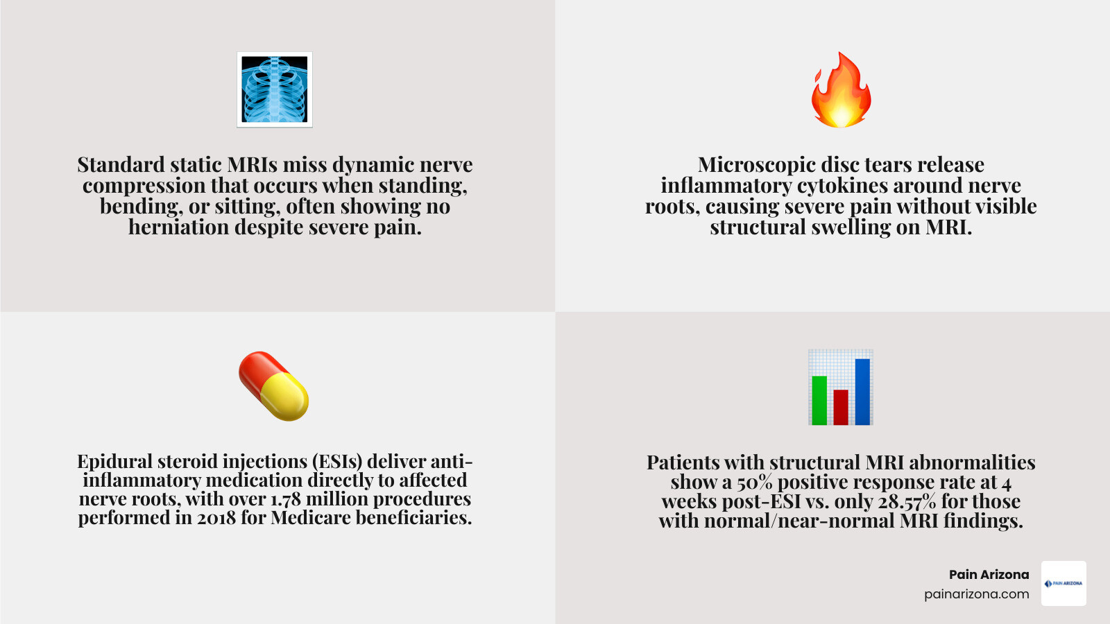
While ESIs deliver powerful anti-inflammatory medication directly around affected nerve roots, recent clinical studies illustrate a clear outcome gap based on MRI findings:
| Clinical Variable | Structural MRI Abnormalities | Normal / Near-Normal MRI |
|---|---|---|
| 4-Week Positive Response Rate | 50.00% | 28.57% |
| Typical Onset of Significant Relief | Within 4 days | Within 4 days (if responsive) |
| Primary Mechanism | Mechanical decompression & anti-inflammatory | Pure anti-inflammatory / Diagnostic response |
Clinical research demonstrates that over 90% of patients who achieve meaningful relief (>50% reduction in pain) from a lumbar ESI report this benefit by day four post-procedure. Evaluating whether a treatment provides rapid relief is crucial when determining whether Is There a Most Effective Treatment Protocol for Lumbosacral Radiculopathies.
ESI Candidacy in Lumbar Radiculopathy Interventional Pain Management Scottsdale
Given the statistical gap in outcomes between clear structural imaging and normal imaging, selecting the right candidates requires meticulous clinical judgment and expert evaluation. In our practice, we exercise careful discretion—similar to top spine surgeons—avoiding repeat procedures if an initial injection offers no measurable analgesic or functional benefit.
Ideal candidates present with:
- Clear neurological physical exam findings matching a distinct nerve root distribution (e.g., dermatomal sensory loss or diminished deep tendon reflexes).
- Failure to progress after 4 to 6 weeks of structured physical therapy and oral anti-inflammatory treatments.
- Concordant pain presentation, even if structural compression on static imaging appears minor.
When an initial ESI yields no response in a normal-MRI patient, non-responder protocols prompt us to pause, re-evaluate, and seek alternative pain generators rather than routinely repeating ineffective injections. Finding appropriate care involves exploring Finding the Best Lumbar Radiculopathy and Sciatica Treatment in Phoenix.
Differential Diagnoses and Shared Decision-Making

When lumbar spine MRIs show clear nerve root spaces despite severe leg discomfort, interventional physicians must carefully evaluate peripheral conditions that closely mimic radiculopathy.
Common non-spinal and peripheral mimics include:
- Piriformis Syndrome: Spasm or inflammation of the piriformis muscle compressing the sciatic nerve in the deep buttock region.
- Common Peroneal Nerve Entrapment: Nerve compression near the fibular head causing lateral leg pain and foot drop symptoms.
- Peripheral Neuropathy: Systemic nerve dysfunction (often related to metabolic or diabetic factors) producing stocking-glove sensory changes.
- Hip Osteoarthritis: Intra-articular hip joint degeneration referring pain deep into the groin, anterior thigh, or knee.
- Vascular Insufficiency: Poor lower extremity arterial circulation causing activity-induced leg cramping and pain (claudication).
Investigating these alternatives ensures patients receive targeted treatment rather than unneeded spinal procedures. See our specialized insights on Getting to the Root of Left Leg Radiculopathy and Pain.
Shared Decision-Making and Future Research Directions
At Pain Arizona, we emphasize transparent communication and shared decision-making. When proposing an ESI for a patient with normal spine imaging, we outline realistic expectations, discussing the lower statistical success rate (roughly 28.5% versus 50% in structural cases) so patients can make fully informed choices about their care.
Current research limitations highlight the need for dynamic functional imaging technologies and advanced biochemical markers capable of detecting micro-inflammation. As these diagnostic tools evolve, our ability to identify subtle pain targets will continue to improve, allowing for increasingly customized interventional plans.
Frequently Asked Questions About Lumbar Radiculopathy
How effective are epidural steroid injections if my spine MRI is normal?
While ESIs remain a viable diagnostic and therapeutic option, studies show they have a lower positive response rate (around 28.57%) in patients with completely normal MRIs compared to those with visible structural nerve compression (50%). However, for patients whose pain stems from chemical inflammation without gross anatomical swelling, an ESI can still provide meaningful relief.
How fast do patients notice pain relief after a lumbar ESI?
The therapeutic response to an epidural corticosteroid injection typically occurs quickly. Clinical evidence indicates that over 90% of positive responders notice significant pain reduction (>50% relief) within four days following the procedure.
What alternative conditions mimic lumbar radicular leg pain?
Conditions that frequently mimic lumbar radiculopathy include piriformis syndrome, sacroiliac joint dysfunction, hip joint osteoarthritis, common peroneal nerve entrapment, peripheral neuropathy, and peripheral vascular disease.
Conclusion
Living with radiating lower back and leg pain is challenging, particularly when diagnostic imaging appears unrevealing. At Pain Arizona, our double board-certified specialists excel in navigating these complex clinical scenarios across Scottsdale, Phoenix, Mesa, Gilbert, and Glendale. By blending precise diagnostic evaluations, electrodiagnostic functional testing, and targeted conservative and interventional therapies, we focus on identifying and treating the true root source of your pain.
We accept most major insurance plans—check with our office manager to confirm if your plan is accepted. If you are ready to find real answers and explore personalized pathways to relief, Schedule a Consultation for Lumbar Radiculopathy Relief with our dedicated medical team today.