
Getting to the Root of Left Leg Radiculopathy and Pain
When Your Lower Back Pain Travels Down Your Left Leg
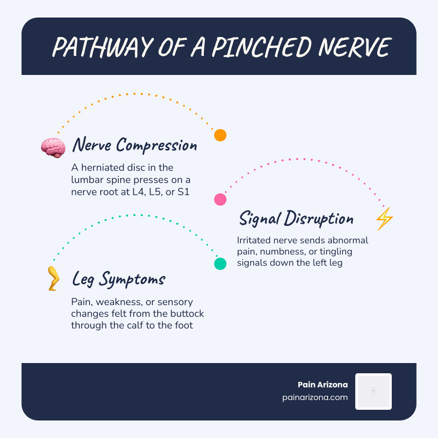
Left leg radiculopathy is a condition where a compressed or irritated nerve root in the lower spine sends pain, numbness, tingling, or weakness down into the left leg — often as far as the foot.
Quick answer: What is left leg radiculopathy?
| Feature | Details |
|---|---|
| What it is | Compression or irritation of a lumbar nerve root causing symptoms in the left leg |
| Common symptoms | Shooting pain, numbness, tingling, weakness down the left leg |
| Most common cause | Herniated disc pressing on a nerve root (L4, L5, or S1) |
| Who it affects | Most common between ages 45–64 |
| Does it go away? | About 90% of cases improve within 3 months with conservative care |
| When to act fast | Loss of bladder/bowel control, sudden severe weakness — seek care immediately |
If you’ve been feeling a sharp, burning, or electric-shock sensation running from your lower back down through your left buttock, thigh, or calf, you’re not alone. Lumbar radiculopathy affects an estimated 3%–5% of the general population, and the left leg is one of the most commonly affected sides.
The pain can range from a dull ache to a jolt that stops you mid-step. It often gets worse when you sit, sneeze, or cough — and it can make everyday life in Phoenix feel exhausting and discouraging, especially when previous treatments haven’t given you real answers.
The good news? Most cases are very treatable — but getting better starts with understanding exactly what’s happening in your spine.
I’m Dr. Nasser, a double board-certified physician in Physical Medicine & Rehabilitation and Pain Medicine, with extensive experience diagnosing and treating left leg radiculopathy through both conservative care and minimally invasive interventional procedures. As Chief Medical Officer at Pain Arizona, I’ve helped thousands of patients across the Greater Phoenix Area find lasting relief by targeting the root cause of their nerve pain — not just masking the symptoms.
Understanding Left Leg Radiculopathy and Its Causes
To understand why your left leg is hurting, we have to look at the anatomy of your lower back. Your lumbar spine is built of stacked bones called vertebrae, cushioned by soft, gel-filled intervertebral discs. Running through the center of these bones is your spinal cord. At each spinal level, nerve roots exit the spinal canal through small bony openings called foramina, traveling down into your hips, thighs, knees, calves, and feet.
When something compromises these small exit pathways on the left side of your spine, the corresponding nerve root becomes pinched or inflamed. This is the physiological basis of left leg radiculopathy.
Several degenerative or traumatic spinal conditions can lead to this localized compression:
- Herniated Discs: The soft inner core of a spinal disc pushes through a tear in its tough outer shell, directly pressing against the adjacent left nerve root.
- Foraminal Stenosis: The natural wear-and-tear of aging can narrow the bony exit holes (foramina), crowding the nerve root.
- Bone Spurs (Osteophytes): Over time, arthritis can cause abnormal bony growths to project into the spinal canal or foraminal spaces, rubbing against the nerve.
- Spondylolisthesis: This occurs when one vertebra slips forward over the one below it, distorting the spinal canal and pinching the exiting nerves.
For a deeper medical dive into how these mechanical issues alter nerve function, you can read the Clinical overview of lumbosacral nerve compression.
How Left Leg Radiculopathy Differs from General Lumbar Issues
It is very common for patients to confuse generalized lower back pain with true radiculopathy. General lumbar issues — like a lumbar muscle strain or facet joint arthritis — typically cause localized, dull, aching pain confined to the lower back or upper buttocks.
In contrast, left leg radiculopathy is characterized by unilateral (one-sided) symptoms that travel along a specific nerve pathway. While you might feel some back pain, the dominant and most disruptive symptoms are felt in your left leg. This is because the compression is localized specifically to the left-exiting nerve roots that merge to form larger peripheral nerves, such as the sciatic nerve. If you want to learn more about how these symptoms present, check out this More info about lumbar radiculopathy symptoms.
Common Spinal Conditions Triggering Left Leg Radiculopathy
Why does the pain prefer one leg over the other? The answer lies in the direction of the spinal structural changes.
Most lumbar disc herniations are “posterolateral.” Because the posterior longitudinal ligament (a strong band of tissue running down the back of the spine) is thickest and strongest in the dead center, spinal discs almost always rupture slightly to one side or the other. When a disc herniates posterolaterally to the left at the L4-L5 or L5-S1 level, it compresses the left traversing or exiting nerve root, leaving the right side completely untouched.
Similarly, ligamentum flavum hypertrophy — a condition where the ligaments inside the spinal canal thicken due to aging — can occur unevenly, narrowing the left side of the canal more than the right. To explore these structural triggers further, refer to this Detailed guide on lumbar radiculopathy.
Nerve Root Mapping: L4, L5, and S1 Patterns
Because each nerve root in your lower back is responsible for a specific map of sensation (dermatome) and muscle movement (myotome) in your leg, we can often tell exactly which nerve is pinched just by listening to your symptoms.
| Nerve Root | Pain & Sensation Pathway (Dermatome) | Muscle Weakness (Myotome) | Affected Reflex |
|---|---|---|---|
| L4 | Front of the thigh, wrapping across the knee to the inner calf | Knee extension (straightening the leg) | Patellar (Knee-jerk) |
| L5 | Outer side of the thigh, lateral calf, and the top of the foot/big toe | Foot dorsiflexion (lifting the foot up) | None (or minimal) |
| S1 | Back of the thigh, back of the calf, outer edge of the foot, and sole | Plantar flexion (pushing down like a gas pedal) | Achilles (Ankle-jerk) |
The L4 Nerve Root and Anterior Thigh Pain
When the L4 nerve root on the left side is compressed, the symptoms primarily affect the front of your leg. You may experience sharp pain or numbness traveling down your anterior thigh and wrapping across your knee.
Because the L4 nerve root helps power the quadriceps muscle, you might notice your left knee feels unstable or “gives out” when you walk or climb stairs. During a physical exam, we will often find a diminished or completely absent left patellar reflex. For an in-depth look at this specific presentation, see our Guide to acute lumbosacral radiculopathy.
The L5 Nerve Root and Foot Drop
The L5 nerve root is the most frequently compressed nerve in the lumbar spine. When irritated on the left, it sends sharp, electric pain down the outer side of your left thigh, running down the lateral calf and wrapping onto the top of your foot, often ending in your big toe.
The L5 nerve root is also responsible for dorsiflexion — the action of pulling your foot and toes upward toward your shin. Severe L5 compression can weaken these muscles, leading to a condition called “foot drop.” If you have foot drop, your left foot may drag on the floor when you walk, forcing you to lift your knee higher to avoid tripping.
Fortunately, there are highly targeted ways to address this. You can read about How to treat foot drop from L5 radiculopathy as well as our Complete guide to L5 radiculopathy treatment.
The S1 Nerve Root and Calf Weakness
An S1 radiculopathy is what most people picture when they think of classic “sciatica.” The S1 nerve root supplies the back of your leg. Compression here sends shooting, hot pain down your left buttock, down the back of your thigh and calf, and into the outer edge or sole of your left foot.
Because S1 controls plantar flexion (the calf muscles used to push off the ground), you might find it difficult to stand up on your left tiptoes. On examination, a pinched S1 nerve root typically results in a diminished or absent Achilles tendon reflex.
How Left Leg Nerve Compression is Diagnosed and Evaluated
Getting you back on your feet starts with an accurate diagnosis. We don’t just treat the pain; we locate the exact point of compression. A comprehensive diagnosis combines your detailed clinical history, a targeted physical exam, advanced imaging, and sometimes specialized nerve testing. To read more about the clinical framework of this evaluation, consult this Scientific review of lumbosacral radiculopathy.
Physical Exam Maneuvers and Clinical Tests
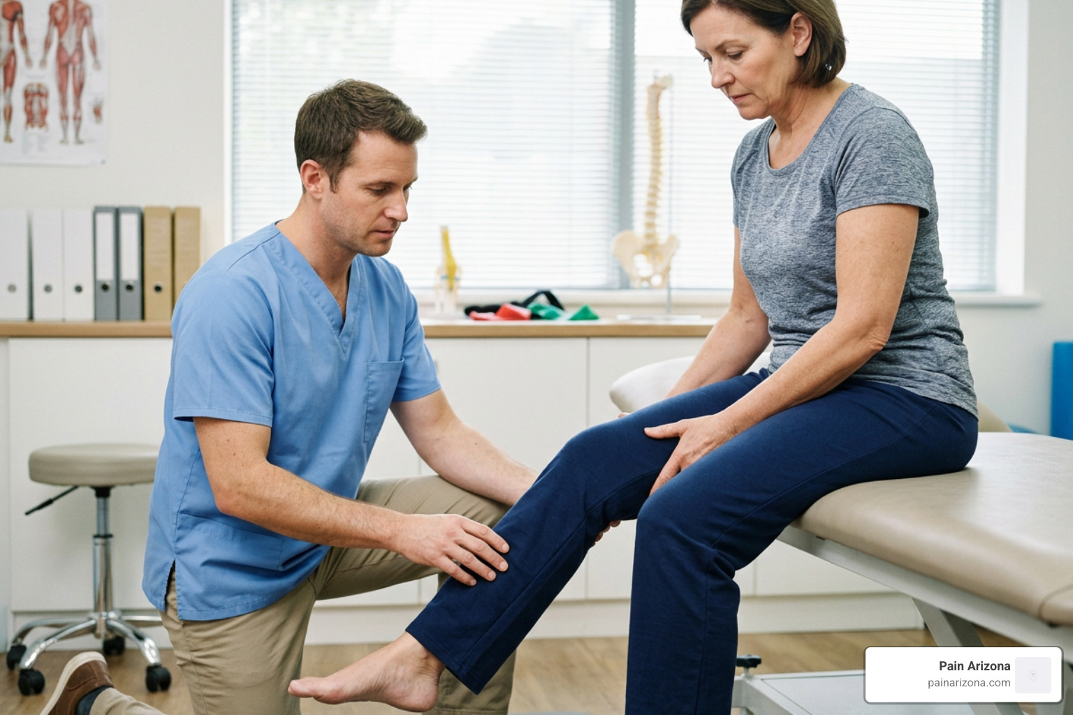
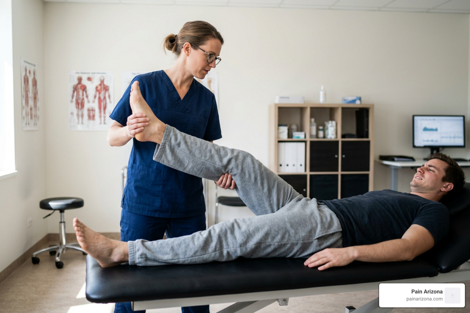
During your physical exam, we perform specific physical maneuvers to see if we can safely reproduce your left leg symptoms.
- The Straight-Leg Raise (Lasegue’s Sign): While you lie flat on your back, we gently lift your straight left leg. If you feel a sharp, shooting pain travel down past your knee between a 30-to-60-degree angle, the test is positive. This indicates that the nerve roots (usually L5 or S1) are inflamed and stretched.
- The Crossed Straight-Leg Raise: We lift your right (unaffected) leg. If lifting your right leg reproduces the shooting pain down your left leg, this is a highly specific sign of a herniated disc on the left.
- The Slump Test: While sitting, you slump forward, chin to chest, and we gently extend your left knee and flex your foot. This puts maximum tension on the spinal cord and nerve roots to identify hidden compression.
Advanced Imaging and Electrodiagnostic Studies (EMG/NCV)
If your symptoms don’t improve after a few weeks, or if you show signs of muscle weakness, we will order a lumbar MRI without contrast. An MRI is the gold standard for spinal imaging, providing highly detailed pictures of your soft tissues, including discs, ligaments, and individual nerve roots.
If you cannot have an MRI (for instance, if you have a pacemaker), a CT myelogram is an excellent alternative.
In complex cases where your MRI shows multiple bulging discs and we need to determine which exact nerve is causing your symptoms, we perform electrodiagnostic studies:
- Nerve Conduction Velocity (NCV): We apply tiny electrical impulses to your leg to measure how fast signals travel down your nerves.
- Needle Electromyography (EMG): We insert a very fine needle electrode into specific muscles in your left leg and lower back to record electrical activity. This helps us differentiate between a pinched nerve in your back and a localized nerve issue in your leg (like peripheral neuropathy).
To learn more about how these tests pinpoint nerve damage, read about the Electrodiagnostic evaluation of radiculopathy.
Red-Flag Symptoms Requiring Urgent Evaluation
While most cases of left leg radiculopathy can be managed safely over time, certain symptoms indicate a medical emergency. If you experience any of the following, seek immediate emergency medical care:
- Saddle Anesthesia: Sudden numbness or a “pins and needles” sensation in your groin, buttocks, inner thighs, or the parts of your body that would touch a saddle.
- Bowel or Bladder Dysfunction: New difficulty urinating, loss of bowel/bladder control, or fecal incontinence.
- Progressive Motor Weakness: Sudden, rapidly worsening weakness in your left leg, such as an inability to lift your foot at all or your leg buckling beneath you.
- Cauda Equina Syndrome: A rare but severe condition where the bundle of nerves at the base of the spinal cord is heavily compressed, requiring emergency surgery to prevent permanent paralysis.
Treatment Options: From Conservative Care to Surgery
At Pain Arizona, our philosophy is always to start with the most conservative, non-invasive treatments first. The human body is remarkably resilient, and about 90% of patients with disc-related nerve compression see significant improvement within 3 months without ever needing surgery. To see how we design these structured recovery plans, explore our guide: Is there an effective treatment protocol for lumbosacral radiculopathies?.
Medications and Non-Invasive Therapies
The initial phase of recovery is all about calming down nerve inflammation and managing your discomfort so you can move comfortably:
- Activity Modification: We don’t recommend absolute bed rest, as inactivity can cause your spinal muscles to stiffen and weaken. Instead, we teach you to temporarily avoid movements that increase pressure in your spine, such as heavy lifting, deep bending, or twisting at the waist.
- Anti-Inflammatory Medications (NSAIDs): Over-the-counter or prescription-strength NSAIDs (like ibuprofen or naproxen) help reduce the chemical inflammation surrounding the pinched nerve root.
- Neuropathic Pain Medications: Medications like gabapentin or pregabalin work specifically on irritated nerve membranes to quiet down the “electric shock” and burning sensations.
- Physical Therapy: Once your acute pain has settled, a physical therapist will guide you through targeted exercises. We highly recommend McKenzie exercises, which focus on “centralizing” your pain — shifting the symptoms out of your left foot and calf and moving them back up into your lower back, where they are much easier to manage.
For a complete breakdown of these non-surgical options, read Everything you need to know about radiculopathy treatment.
Interventional Procedures and Epidural Steroid Injections
If oral medications and physical therapy aren’t providing enough relief, we can deliver powerful anti-inflammatory medicine directly to the source of the problem.
A transforaminal epidural steroid injection (TFESI) is a highly precise, outpatient procedure. Under live X-ray guidance (fluoroscopy), we direct a thin needle into the epidural space, right next to the inflamed left nerve root. We inject a small mixture of local anesthetic and a concentrated corticosteroid. This bathes the nerve, dramatically reducing swelling and interrupting the pain signals.
These injections can provide months of relief, opening a “window of opportunity” for you to participate fully in physical therapy and rebuild your core strength. We also use targeted diagnostic nerve blocks to confirm exactly which spinal level is generating your pain. For more details on these procedures, read What is the solution for leg pain?.
Surgical Decompression Indications
Surgery is rarely the first step, but it becomes necessary under specific circumstances. We typically recommend a surgical consultation if you have:
- Progressive neurological deficits (like worsening leg weakness or foot drop).
- Intolerable, intractable pain that has failed to improve after 6 to 8 weeks of dedicated conservative therapy.
- Clear structural compression on your MRI that correlates perfectly with your physical symptoms.
The most common surgical options are highly effective and minimally invasive:
- Microdiscectomy: A surgeon makes a tiny incision in your lower back and uses a microscope to remove only the small portion of the herniated disc that is pinching the left nerve root.
- Laminectomy: The surgeon removes a small piece of the bony arch of the vertebra (the lamina) to widen the spinal canal and relieve pressure on compressed nerves.
To compare long-term recovery rates between surgical and non-surgical approaches, you can read this comprehensive study on Surgical vs non-surgical outcomes for disc herniation.
Frequently Asked Questions About Unilateral Nerve Pain
How long does it take for unilateral leg pain to heal?
The natural history of left leg radiculopathy is highly favorable. Most patients experience significant relief within 6 to 12 weeks. The body has an amazing ability to naturally resorb herniated disc material over time. However, recovery is a gradual process, and nerve tissues heal slowly. Consistent physical therapy, proper posture, and avoiding re-injury are key to staying on track.
Can a pinched nerve in the lower back cause permanent damage?
If left untreated for many months, severe and constant nerve compression can lead to permanent nerve damage. This is why we closely monitor your muscle strength and reflexes. While sensory changes like mild numbness can take a long time to fully resolve, progressive motor weakness (like a worsening foot drop) requires prompt medical intervention to ensure the nerve can regenerate successfully.
What exercises should I avoid during recovery?
During your recovery, you should avoid any exercises or movements that put high compressive loads on your spinal discs or force your spine into extreme ranges of motion. Skip heavy squats, deadlifts, sit-ups, deep forward bends (like touching your toes), and high-impact activities like running or jumping. Instead, focus on low-impact cardiovascular exercises like walking or swimming, alongside core-stabilizing movements like the “bird-dog” or “plank” under the guidance of a professional.
Conclusion
Living with left leg radiculopathy can feel overwhelming, but you don’t have to navigate this journey alone. At Pain Arizona, our double board-certified physicians specialize in comprehensive chronic pain management. We serve patients across the Greater Phoenix Area — with state-of-the-art clinics in Phoenix, Mesa, Gilbert, Scottsdale, and Glendale.
Our team is dedicated to creating personalized treatment plans tailored to your unique lifestyle and recovery goals. We believe in compassionate care, utilizing the latest in evidence-based, minimally invasive medicine to help you reclaim your mobility and live pain-free.
We accept most insurance plans. Check with our office manager to see if your plan is accepted.
If you are ready to get to the root of your left leg pain and find lasting relief, take the first step today and Schedule a consultation for lumbar radiculopathy relief with our team.