
A Comprehensive Guide to Compression Fracture Causes
When Your Spine Gives Way: Understanding Compression Fractures
A compression fracture is a break in one of the vertebrae — the bones that stack together to form your spine — where the bone collapses under pressure. Here’s what you need to know at a glance:
| Question | Quick Answer |
|---|---|
| What is it? | A vertebra cracks and collapses, most often in the mid or lower back |
| Most common cause | Osteoporosis (weakened bones) |
| Who is most at risk? | Women over 50, adults over 80, trauma patients |
| Key symptoms | Sudden back pain, loss of height, hunched posture |
| Does it heal? | Often yes — pain typically eases within 4 weeks, with full healing around 12 weeks |
| Treatment options | Bracing, medication, physical therapy, or minimally invasive procedures |
Every year, somewhere between 1 and 1.5 million compression fractures occur in the United States alone. Many happen during ordinary moments — stepping out of a car, sneezing, or simply rolling over in bed. In fact, studies suggest that up to 30% of fractures in people with severe osteoporosis are triggered by something as minor as turning in bed. What makes this condition especially tricky is that roughly two-thirds of people with a compression fracture don’t even know they have one — the pain is absent or so gradual that it gets written off as ordinary back pain.
That distinction matters. A compression fracture isn’t just a bad backache. Left unrecognized or untreated, it can lead to permanent posture changes, lost height, chronic pain, and in serious cases, nerve damage.
I’m Dr. Nasser — a double board-certified PM&R and pain medicine physician with extensive experience diagnosing and treating spinal conditions, including vertebral compression fractures, using minimally invasive and evidence-based approaches. In this guide, I’ll walk you through everything you need to understand about this condition, from its root causes to your real treatment options.
What is a Vertebral Compression Fracture?
To understand a compression fracture, it helps to look at the anatomy of your backbone. Your spinal column is built from 33 individual bones called vertebrae. The solid, drum-shaped front portion of each bone is called the vertebral body. These bodies stack on top of each other, separated by rubbery intervertebral discs that act as shock absorbers.
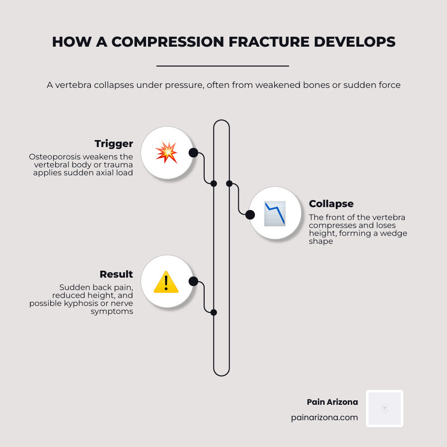
When a vertebral body suffers a compression fracture, this drum-shaped structure physically collapses under load. The bone develops tiny cracks, weakens, and squashes down.
While a compression fracture can happen anywhere in the spine, it is most common in the middle back (the thoracic spine) and the lower back (the lumbar spine). Specifically, the area where these two regions meet — known as the thoracolumbar junction (spanning from the T12 to the L2 vertebrae) — is the most vulnerable. Studies show that 60% to 75% of all vertebral compression fractures occur at this junction, with another 30% occurring in the L2 to L5 region.
To learn more about the fundamental anatomy and mechanics of this injury, explore our Vertebral Compression Fracture Guide or read this clinical clinical overview of vertebral compression fractures.
How a Compression Fracture Differs from Other Spinal Fractures
Not all broken backs are the same. A simple fracture in another bone might mean a clean break right through the middle. In the spine, however, a compression fracture is characterized by a crushing mechanism.
When axial loading — downward pressure combined with the natural forward bending of the spine — exceeds the strength of the vertebral bone, the bone tissue is squashed. Unlike high-impact fractures that can completely shatter the spinal column and threaten spinal stability, a typical osteoporotic compression fracture only affects the front (anterior) part of the vertebra. This leaves the back (posterior) columns intact, meaning the spine usually remains stable, and the spinal cord remains safe from immediate compression.

The Three Main Types of Compression Fracture
Medical professionals classify these fractures to determine the best treatment pathway. The two primary grading systems are the Genant classification (which measures the severity of height loss) and the AO Spine classification (which looks at the fracture’s structural pattern).
There are three main physical shapes a compression fracture can take:
- Wedge Fracture: This is the most common type, accounting for more than half of all cases. The front (anterior) part of the vertebral body collapses, while the back (posterior) part remains at its normal height. This creates a wedge-shaped bone, which tilts the spine forward.
- Crush Fracture: In a crush fracture, the entire vertebral body collapses uniformly. Both the front and back of the bone lose height, which can severely impact overall spinal height.
- Burst Fracture: This is a more severe, unstable fracture. The vertebra loses height in both the front and back, and the bone breaks into multiple fragments that can spread out in several directions. If these fragments push backward into the spinal canal (a phenomenon known as retropulsion), they can press against the spinal cord or nerve roots, causing serious neurological issues.
| Fracture Type | Structural Mechanism | Clinical Significance |
|---|---|---|
| Wedge | Anterior collapse only; posterior height preserved. | Most common; leads to a forward-leaning posture (kyphosis). |
| Crush | Entire vertebral body collapses uniformly. | Causes significant loss of overall height and back pain. |
| Burst | Multi-directional breakage; posterior wall disrupted. | High risk of spinal instability and nerve damage; requires urgent evaluation. |
Causes, Symptoms, and Diagnosis of Vertebral Fractures
A healthy vertebral body is remarkably strong. For it to collapse, either the bone itself must be significantly weakened, or the force applied to it must be incredibly intense.
Osteoporosis and Bone Density Loss
By far, the leading cause of compression fractures is osteoporosis, a systemic metabolic bone disease where the body loses too much bone, makes too little bone, or both. This leaves the internal “honeycomb” structure of the bone thin and brittle. A milder form of bone thinning is known as osteopenia.
Postmenopausal women are at the highest risk for osteoporotic fractures due to the drop in estrogen, a hormone that protects bone density. Additionally, nutritional deficiencies — specifically calcium deficiency and vitamin D deficiency — prevent the body from rebuilding strong bone.
According to population studies, the annual incidence of vertebral compression fractures is 10.7 per 1000 women and 5.7 per 1000 men. By the time they reach 80 years of age, 40% of all women will have sustained at least one compression fracture. To dive deeper into the relationship between bone density and spinal health, you can read the comprehensive research on osteoporotic spinal fractures.
High-Impact Trauma and Accidents
In younger patients with healthy bones, a compression fracture is almost always the result of high-energy trauma.
- Motor Vehicle Collisions: In younger demographics, about 50% of spine fractures are caused by car crashes.
- Falls from Height: Another 25% are caused by falls, such as falling from a ladder or scaffolding.
- Sports Injuries: High-impact sports can also generate an extreme axial load that crushes the vertebral bodies.
An interesting clinical fact: about 10% of patients who suffer a calcaneus (heel bone) fracture from a fall from height also have an associated thoracolumbar fracture. This is because the massive force of landing on one’s feet travels straight up the skeletal system into the spine.
Spinal Malignancy and Pathologic Fractures
When a vertebra is weakened by an underlying medical condition like cancer, it is called a pathologic fracture.
Metastatic cancer (cancer that has spread to the spine from other organs like the breasts, lungs, or prostate) and multiple myeloma (a cancer of the plasma cells in the bone marrow) can infiltrate the vertebral body. As tumor cells replace healthy bone tissue, the vertebra becomes structurally hollow and can collapse under normal daily loads.
Clinically, if a patient presents with a solitary compression fracture located above the T4 vertebra without a history of severe trauma, we highly suspect a neoplastic (cancerous) cause, as standard osteoporotic fractures rarely occur that high up the spine. For a highly detailed clinical perspective, refer to the professional guidelines on pathologic fractures.
Recognizing the Signs of Vertebral Collapse
Because many of these fractures happen gradually or present with mild symptoms, knowing what to look for is essential. Common signs include:
- Sudden Sharp Back Pain: Often triggered by a minor action like bending over or coughing. The pain typically worsens when standing or walking and improves when lying flat on your back.
- Loss of Height: Over time, as multiple vertebrae collapse, a person may lose several inches of height.
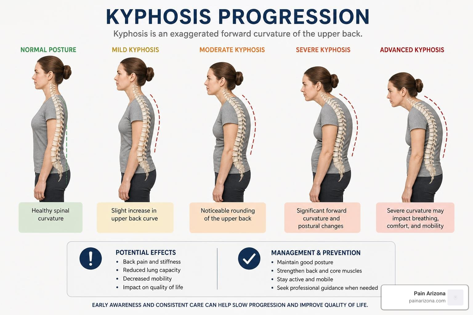
- Kyphosis (Dowager’s Hump): When multiple wedge fractures occur, the spine curves forward, creating a rounded hump in the upper back.
- Muscle Spasms: The muscles surrounding the injured area work overtime to stabilize the spine, leading to painful spasms.
- Neurological Symptoms: If bone fragments press on nerves, you may experience numbness, tingling, radiating pain down the legs, or muscle weakness.
Diagnostic Exams and Imaging Modalities
If you visit our offices in the Greater Phoenix Area, we use a combination of physical exams and advanced imaging to pinpoint the issue.
Physical Examination
During your physical, we may perform specific provocative tests:
- Closed-Fist Percussion Test: Gently tapping along your spine with a closed fist. If a specific vertebra is fractured, this causes sharp, localized discomfort (this test has an 88% sensitivity and 90% specificity).
- Supine Sign Test: Asking you to lie flat on your back with only a pillow. Patients with acute fractures often cannot tolerate this position due to severe pain (with 81% sensitivity and 93% specificity).
- Back Pain-Inducing Test: A movement-based assessment to see if spinal rotation or flexion reproduces the pain.
Imaging Modalities
- Spinal Radiographs (X-rays): The initial step to confirm a fracture. A loss of vertebral height of 20% or more compared to surrounding vertebrae is the standard diagnostic benchmark.
- MRI Scan: The gold standard for determining if a fracture is acute (new) or chronic (old). It shows bone marrow edema (swelling) and reveals if nerves are being compressed.
- CT Scan: Excellent for viewing bone details and evaluating whether a fracture is stable or if bone fragments have broken off into the spinal canal.
- DEXA Scan: A bone density test used to diagnose osteoporosis and calculate future fracture risks.
For a technical breakdown of how radiologists identify these injuries, you can view the radiographic features of spinal fractures.
Treatment, Recovery, and Prevention Strategies
Fortunately, you have many options when it comes to managing a compression fracture. The right path depends on your pain levels, bone health, and whether the fracture is stable.
Non-Surgical and Conservative Management
Most stable compression fractures can heal on their own over several months with conservative care.
- Activity Modification: We recommend avoiding heavy lifting, twisting, and high-impact activities. However, prolonged bed rest is discouraged, as it can worsen bone loss and lead to muscle wasting.
- Pain Medications: Short-term use of over-the-counter pain relievers like acetaminophen or nonsteroidal anti-inflammatory drugs (NSAIDs) can help manage pain.
- Calcitonin: A prescription medication that can help reduce bone pain during the acute healing phase.
- Back Bracing: Wearing a supportive back brace (like a TLSO brace) for 4 to 12 weeks can limit spinal movement, reduce pain, and assist with healing. However, bracing is typically limited to the acute phase (under 8 weeks) to prevent core muscle weakness.
- Physical Therapy: Once the initial pain decreases, targeted physical therapy is vital to strengthen the core and back muscles that support your spine.
To explore conservative treatment options further, check out our guides on Lower Back Pain Treatments and our Back Pain Treatment Complete Guide.
Minimally Invasive and Surgical Interventions
If conservative therapies do not relieve your pain after 6 weeks, or if you have severe, debilitating pain, minimally invasive procedures can provide rapid relief.
- Kyphoplasty: A highly effective, outpatient procedure. A small balloon is inserted into the collapsed vertebra and gently inflated to restore the bone’s height. The balloon is then deflated, and the resulting space is filled with fast-drying medical bone cement to stabilize the fracture. Most patients experience significant pain relief within 24 to 48 hours.
- Vertebroplasty: Similar to kyphoplasty, but bone cement is injected directly into the fractured vertebra without using a balloon first.
- Spinal Fusion: Reserved for rare, unstable fractures (like severe burst fractures) or cases with spinal cord compression. This major surgery permanently joins two or more vertebrae together to stabilize the spine.
To learn more about these options, read about our approaches to Minimally Invasive Spine Surgery and what to expect regarding Back Surgery Recovery Time.
Expected Recovery Timeline and Prognosis
For most patients, the healing journey follows a predictable path:
- Weeks 1 to 4: Pain is typically at its highest but gradually begins to decrease.
- Weeks 4 to 12: The bone continues to knit back together. Most patients experience complete healing and a return to normal daily activities by week 12.
- Long-Term: If the underlying osteoporosis is not treated, the risk of sustaining another fracture is five times higher.
While the fracture itself heals, studies show that older adults who suffer a compression fracture have lower long-term survival rates (53.9% at 3 years, 30.9% at 5 years, and 10.5% at 7 years). This is not usually due to the fracture itself, but because a fracture often indicates severe underlying osteoporosis, frailty, and an increased risk of future debilitating falls. For more patient-focused recovery insights, check out this patient guide to spinal fractures.
Long-Term Prevention and Bone Health
Preventing your first (or next) compression fracture comes down to protecting your bone density and minimizing fall risks.
- Nutrition: Ensure you receive adequate calcium (1,200 mg daily) and vitamin D (800 to 1,000 IU daily) through diet or supplements.
- Exercise: Engage in regular weight-bearing exercises (like walking) and resistance training to stimulate bone growth.
- Lifestyle Changes: Quitting smoking and limiting alcohol intake are critical, as both nicotine and excessive alcohol accelerate bone loss.
- Fall Proofing: Remove tripping hazards at home, install grab bars in bathrooms, and ensure your living spaces are well-lit.
- Medical Management: Work with a specialist to evaluate your bone density and discuss prescription bone-building or bone-preserving medications (like bisphosphonates).
For further clinical guidance on preventing fractures, review the AAFP recommendations on fracture prevention and learn how to choose the right partner for your care in our guide on Spine and Pain Management: How to Choose Your Specialist.
Frequently Asked Questions about Spinal Fractures
Can a collapsed vertebra heal on its own?
Yes. Many stable compression fractures heal on their own over a period of about 12 weeks. This process requires conservative management, which may include wearing a specialized back brace, modifying your physical activities, and taking pain-relief medications. However, it is essential to have any suspected fracture evaluated by a medical professional to ensure it is stable and healing correctly.
How serious is a spinal collapse?
A compression fracture can range from mild to very serious. While many are stable and cause minimal symptoms, severe or untreated fractures can lead to:
- Chronic, debilitating back pain.
- Significant height loss and a hunched posture (kyphosis), which can compress internal organs and make breathing difficult.
- Spinal cord or nerve compression, leading to numbness, weakness, or loss of bowel and bladder control (which is a medical emergency).
What is the life expectancy after a vertebral break?
A compression fracture does not directly shorten your life expectancy. However, because these fractures are highly correlated with advanced age and severe osteoporosis, they serve as a major warning sign for general physical frailty. Patients who experience a fracture are at a much higher risk for subsequent falls and secondary health complications. Managing your bone health and working with a specialist can help you maintain a normal, active life expectancy.
Conclusion
A compression fracture can feel overwhelming, but you do not have to live with debilitating back pain or watch your posture change. At Pain Arizona, we specialize in comprehensive, compassionate chronic pain management across the Greater Phoenix Area — with convenient locations in Phoenix, Mesa, Gilbert, Scottsdale, and Glendale.
Our double board-certified physicians focus on identifying the root cause of your pain and offering evidence-based, minimally invasive treatments (like kyphoplasty) to help you regain your mobility and quality of life. We accept most major insurance plans; please check with our office manager to confirm that your specific plan is accepted.
If you or a loved one is struggling with sudden or chronic back pain, do not wait. Schedule a consultation for vertebral compression fracture care with us today and take your first step toward recovery.