
Effective Pain Management Options for Osteoporosis and Chronic Fractures
A Practical Start to Osteoporosis Fracture Pain Management

Effective osteoporosis fracture pain management starts with finding the fracture and matching treatment to the pain level. Seek prompt medical care for sudden severe back, hip, or wrist pain after a minor fall or everyday movement. Then use a plan that combines safe pain relief, brief support such as bracing when appropriate, early guided movement, and osteoporosis treatment to lower the chance of another fracture.
For a painful vertebral compression fracture, pain is often strongest at the start and commonly improves within 3 to 6 weeks. But do not assume ongoing pain is “just aging.” Persistent pain can come from changes in posture, muscle strain, nerve irritation, or another untreated fracture.
Pain control should help you safely stand, walk, sleep, and begin recovery without creating new problems such as sedation, constipation, confusion, or falls. This is especially important for older adults and anyone taking several medicines.
I’m Dr. Nasser, a double board-certified PM&R and pain medicine physician with advanced training in minimally invasive spine procedures, rehabilitation, and complex pain care. At Pain Arizona, I approach osteoporosis fracture pain management by identifying the pain source, protecting function, and building a personalized plan that reduces reliance on medication when safer, lasting options are available.
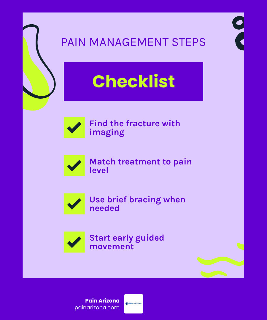
Osteoporosis fracture pain management terms simplified:
Understanding Osteoporosis Pain: Acute Fractures vs. Chronic Mechanical Strain
Osteoporosis is often termed a “silent disease” because bone loss alone causes no physical symptoms. Bone mineral density naturally declines beginning around age 40, and a diagnosis is established when a dual-energy X-ray absorptiometry (DXA) scan reveals a T-score of -2.5 standard deviations or lower. Pain only emerges when fragile skeletal microarchitecture yields under routine physiological loads. Understanding the biological distinction between acute fracture trauma and subsequent chronic mechanical dysfunction is critical for providing lasting relief.
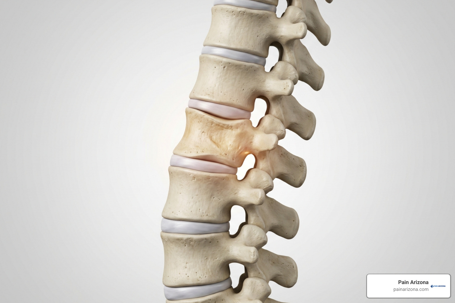
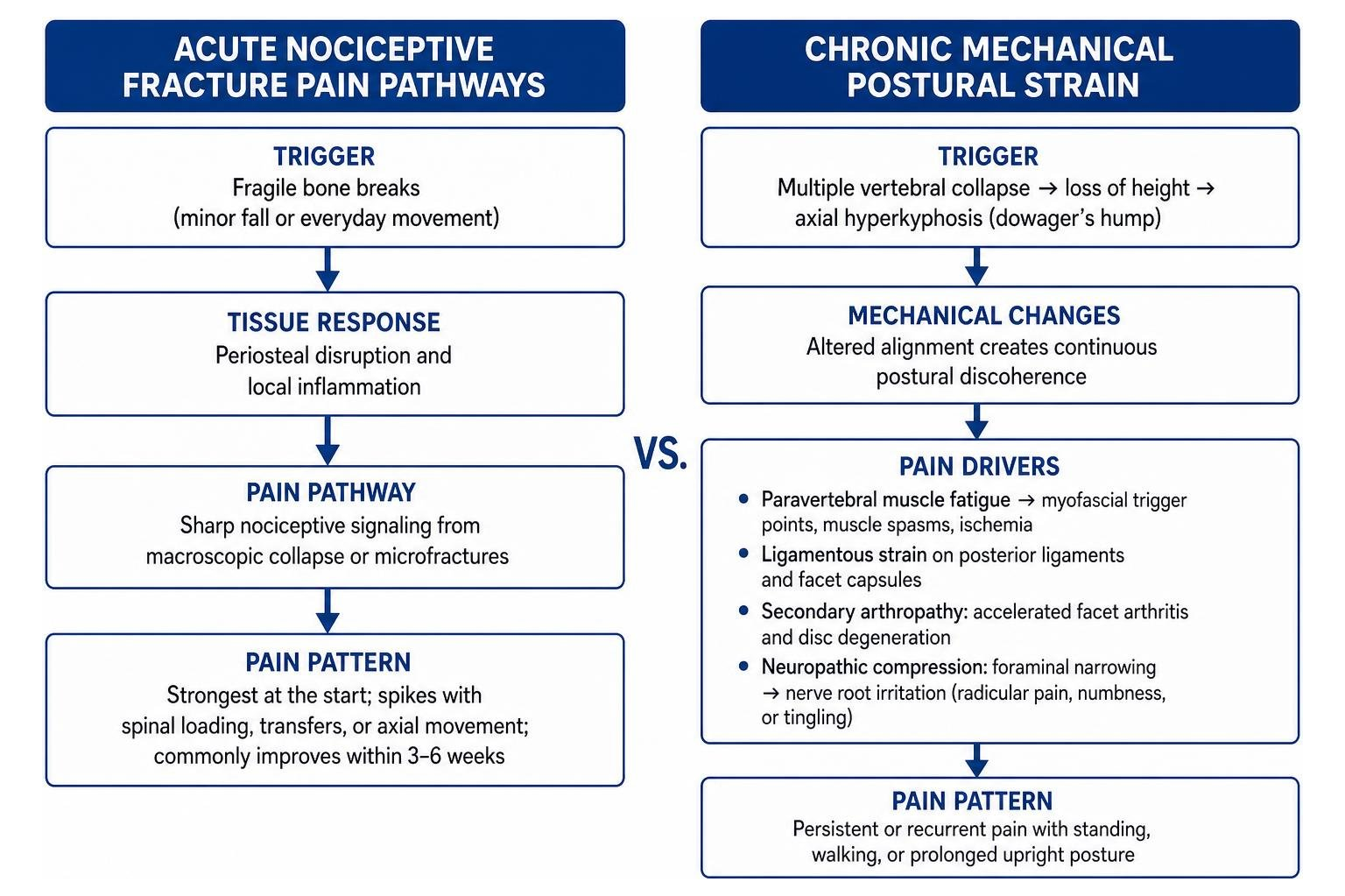
Acute pain stems from sharp nociceptive signaling triggered by macroscopic structural collapse or microscopic trabecular microfractures. When a weakened bone breaks—such as when lifting a grocery bag or stepping off a curb—periosteal disruption and local inflammation generate sharp, immediate distress that spikes during spinal loading, transfers, or axial movement.
Conversely, chronic pain in osteoporosis rarely reflects ongoing bone breakage alone. As multiple vertebral bodies collapse, the anterior spinal column loses vertical height, driving the spine into progressive axial hyperkyphosis (often referred to as a dowager’s hump). This altered biomechanical alignment creates continuous postural discoherence:
- Paravertebral Muscle Fatigue: Posterior back extensor muscles must maintain constant, tonic contraction just to keep the torso upright, causing severe myofascial trigger points, chronic muscle spasms, and localized ischemia.
- Ligamentous Strain: Anterior spinal shortening places persistent tension on the posterior interspinous ligaments and facet joint capsules.
- Secondary Arthropathy: Shifted center-of-gravity forces accelerate degenerative facet joint arthritis and intervertebral disc degeneration.
- Neuropathic Compression: Narrowing neural foramina can pinch exiting spinal nerve roots, producing radicular pain, numbness, or tingling traveling across the ribcage or down the lower extremities.
Clinical Pain Assessment and the Reverse WHO Analgesic Ladder
Accurately capturing pain intensity requires validated tools like the 11-point Numeric Rating Scale (NRS) or the Visual Analogue Scale (VAS). Because pain catastrophizing, depression, and fear of falling can amplify nociceptive processing and elevate the risk of transition into chronic disabling pain, our clinical evaluations screen for psychological distress alongside physical symptoms.
Traditional pain management follows the World Health Organization (WHO) analgesic ladder, which starts with mild non-opioid medications and slowly steps upward as pain persists. For an acute osteoporotic fragility fracture, this upward model often fails. Acute fracture pain peaks immediately upon structural failure. Applying a step-up model leaves vulnerable patients immobilized and in agony during the crucial first days of injury.
According to authoritative clinical insights like the ROS Guidance for the management of symptomatic vertebral fragility fractures, a reverse WHO analgesic ladder is often far more clinically effective. Physicians provide robust, rapid pain control during the initial acute window, enabling prompt out-of-bed mobilization, and then systematically down-titrate medications as the bone stabilizes and inflammatory cytokines subside over 3 to 6 weeks.
Comprehensive Guidelines for Osteoporosis Fracture Pain Management
Managing fragility fractures requires a comprehensive balance between immediate analgesic potency and patient safety. Because chronic pain affects 41% of individuals aged 65–75 and up to 55% of those over 85, our primary clinical goal is preventing prolonged bed rest. Extended immobilization rapidly accelerates bone mineral loss, triggers profound sarcopenic muscle wasting, and increases the risks of deep vein thrombosis and pneumonia.
| Treatment Phase | Primary Clinical Goals | First-Line Strategies | Key Precautions & Avoidance |
|---|---|---|---|
| Acute Phase (Weeks 0–6) | Rapid nociceptive suppression, early safe mobilization, fracture stabilization | Scheduled paracetamol, short-term NSAIDs/targeted nerve blocks, short-term rescue opioids, dynamic bracing | Prolonged bed rest (>48 hrs), over-sedation, unmonitored NSAID toxicity, heavy opioid reliance |
| Subacute Phase (Weeks 6–12) | Structural remodeling, weaning analgesics, progressive extensor loading | Osteoanabolic agents (e.g., teriparatide), active physical therapy, isometric core strengthening | Trunk flexion/twisting, sudden heavy lifting, permanent rigid brace dependency |
| Chronic Phase (Month 3+) | Postural realignment, myofascial relief, secondary fracture prevention | Mind-body therapy, back extensor conditioning, antiresorptives, Fracture Liaison Services (FLS) | High-impact spinal loading, unaddressed fall hazards, untreated skeletal bone loss |
Pharmacological Approaches to Osteoporosis Fracture Pain Management
Medication regimens must be carefully selected to avoid exacerbating common geriatric comorbidities:
- Scheduled Paracetamol (Acetaminophen): Acts as the foundational non-opioid baseline analgesic. While gentle on the gastrointestinal tract, total daily doses must be carefully tracked to prevent hepatic strain, particularly in frail older adults.
- Non-Steroidal Anti-Inflammatory Drugs (NSAIDs): As outlined in a detailed systematic review of pharmacological options for pain control in vertebral fragility fractures, NSAIDs provide significant short-term relief for movement-related pain. However, their systemic use must be brief and closely monitored due to risks of gastrointestinal ulceration, worsening renal function, cardiovascular strain, and potential interference with early bone healing.
- Short-Term Rescue Opioids: Weak opioids (such as codeine or tramadol) or carefully dosed strong opioids are restricted to severe breakthrough pain to facilitate transfers and physical therapy. Opioids carry high rates of adverse events—approximately 50% of users experience side effects such as severe constipation, cognitive clouding, urinary retention, and increased fall risks, leading over 20% to discontinue therapy. Co-prescribing prophylactic bowel regimens is mandatory.
- Neuropathic Adjuvants: When a compressed vertebra impinges on an exiting nerve root, gabapentinoids (gabapentin, pregabalin) or low-dose tricyclic antidepressants help dampen neuropathic firing and radicular distress.
- Calcitonin: Though historically utilized for short-term acute bone pain, calcitonin is generally restricted due to weak long-term efficacy and potential regulatory safety concerns regarding chronic use.
Bone-Targeted and Anabolic Therapies for Fracture Healing and Analgesia
Modern osteoporosis medications do far more than slowly accumulate bone density; several possess direct, measurable analgesic properties. By suppressing osteoclast-mediated bone resorption, inhibiting inflammatory cytokines (such as tumor necrosis factor-alpha), and downregulating pain-signaling neuropeptides like substance P and calcitonin gene-related peptide (CGRP), these agents actively relieve skeletal pain.
- Osteoanabolic Agents (Teriparatide): Daily or weekly subcutaneous teriparatide (a parathyroid hormone analog) stimulates active osteoblastic bone formation. A major 2024 network meta-analysis published in JAMA Network Open on conservative treatments for acute painful vertebral compression fractures confirmed that teriparatide demonstrates superior long-term pain reduction compared to standard antiresorptives, while driving progressive improvements in physical function over 18 months of care.
- Intravenous Bisphosphonates: Agents such as intravenous pamidronate and zoledronic acid have demonstrated significant reductions in acute vertebral pain within 48 to 72 hours of infusion, stabilizing trabecular microdamage and damping inflammatory bone turnover.
- RANKL Inhibitors (Denosumab): Administered every six months, denosumab significantly reduces spinal pain indices and lowers pain catastrophizing scores by rapidly shutting down accelerated osteoclastic bone resorption.
Interventional and Surgical Options for Spinal and Hip Fragility Fractures
When non-operative therapies fail to control pain or when severe structural collapse prevents a patient from standing, minimally invasive interventions offer rapid stabilization. For patients navigating spinal trauma, reviewing an in-depth vertebral compression fracture guide can clarify structural anatomy and modern interventional pathways.
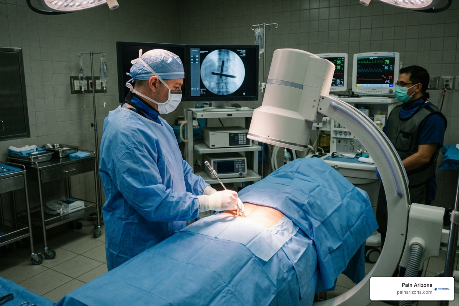
Vertebral Augmentation: Kyphoplasty and Vertebroplasty
Vertebral augmentation procedures are specifically designed to treat painful, unhealed compression fractures with proven bone marrow edema.
- Percutaneous Vertebroplasty: High-viscosity polymethylmethacrylate (PMMA) bone cement is injected directly into the fractured vertebral body under real-time fluoroscopic guidance, stabilizing micro-instability and cementing micro-fracture lines.
- Balloon Kyphoplasty: An inflatable balloon tamp is inserted percutaneously into the collapsed vertebral body and expanded to create a controlled cavity while partially restoring lost vertebral height and reducing kyphotic deformity. The void is then filled with PMMA cement under low pressure, minimizing the risk of cement leakage.
Clinical trials demonstrate that vertebral augmentation is most effective when performed within an early 8-week window from fracture onset. In appropriately selected patients, it provides immediate structural stabilization, allowing them to stand and walk with markedly reduced pain within hours. If you are struggling with severe spine pain in the East Valley or Central Arizona, discovering options for kyphoplasty in Phoenix can open the door to lasting mechanical relief.
Regional Nerve Blocks and Hip Fracture Care
Hip fractures account for up to 51% of fragility-related disability in women and 24% in men. In emergency and perioperative settings, pain management is critical to prevent delirium and facilitate surgical repair.
According to the PROSPECT clinical guideline recommendations published in Acta Anaesthesiologica Belgica, single-shot peripheral regional nerve blocks—specifically Fascia Iliaca Compartment Blocks (FICB) and Femoral Nerve Blocks (FNB)—are first-line analgesic standards. Delivering targeted local anesthetic around these neural pathways reduces systemic opioid requirements by up to 98%, dramatically diminishes perioperative delirium, allows comfortable patient positioning for spinal anesthesia, and substantially reduces the incidence of chronic postsurgical pain (which otherwise affects up to 60% of hip fracture patients at 3 months).
Physical Rehabilitation, Mind-Body Therapies, and Bracing Protocols
Lasting functional recovery requires re-educating the muscular system to support altered skeletal mechanics. To safeguard the spine during early healing, choosing the top braces for lumbar compression fracture recovery can stabilize the injury while helping patients maintain upright posture.
Non-Pharmacological Strategies in Osteoporosis Fracture Pain Management
A structured rehabilitation program must be carefully calibrated to the patient’s structural tolerance:
- Bracing Protocol & Weaning: Dynamic, active semirigid spinal orthoses provide sensory feedback and unload fractured anterior vertebral columns during early ambulation. However, braces must be progressively weaned between 4 to 8 weeks to avoid inducing profound paraspinal muscle deconditioning and weakness.
- Targeted Physical Therapy: Formal physical therapy should begin between 4 to 12 weeks post-injury once acute inflammation resolves. Rehabilitation focuses on isometric back extensor endurance, scapular retractor strengthening, hip abductor stability, and balance training.
- Strict Biomechanical Precautions: Exercises involving loaded spinal flexion (such as traditional sit-ups, toe touches, or deep forward bending) and aggressive axial rotational twisting must be strictly avoided, as flexion places massive compressive loads on compromised vertebral bodies and dramatically increases secondary fracture risk.
- Modalities: Superficial heat application eases tight paraspinal muscle contractures, cold therapy dampens acute inflammatory flares, and specialized postural taping across the thoracic spine enhances proprioceptive feedback. Gentle practices like modified yoga or Pilates emphasize spinal elongation and core stability without forward flexion.
Mind-Body Modalities and Fracture Liaison Services (FLS)
Because chronic pain involves neural amplification and emotional distress, psychological therapies play an essential role in comprehensive care:
- Cognitive-Behavioral Therapy (CBT): Helps patients reframe pain catastrophizing, unlearn fear-avoidance behaviors, and build healthy coping strategies for daily movement.
- Mindfulness & Biofeedback: Mindfulness-based stress reduction (MBSR) and biofeedback teach patients voluntary autonomic control over compensatory muscle tension, lowering systemic stress hormones and dampening central nervous system pain amplification.
- Fracture Liaison Services (FLS): A single vertebral fracture increases the risk of a subsequent spinal fracture five-fold within the first year and nearly triples hip fracture risk. As highlighted by systematic reviews in Pain, Joints, Spine, centralized Fracture Liaison Services coordinate orthopedic care, DXA bone density testing, fall-prevention assessments, and secondary medical therapies to prevent debilitating secondary fractures.
Frequently Asked Questions about Osteoporosis Fracture Pain Management
How long does pain usually last after an osteoporotic vertebral fracture?
Acute fracture pain is typically most severe during the first 7 to 14 days, with approximately two-thirds of patients experiencing substantial spontaneous bone consolidation and pain resolution within 3 to 6 weeks. However, if pain persists beyond 6 to 8 weeks, it usually indicates fracture non-union (pseudarthrosis), unhealed bone marrow edema, or chronic mechanical back pain caused by altered spinal curvature and paraspinal muscle fatigue.
Are back braces recommended for long-term osteoporotic fracture pain relief?
No. While rigid or semirigid back braces are valuable tools for stabilizing the spine and easing movement-related pain during the initial 4 to 8 weeks following a fracture, long-term reliance is counterproductive. Extended brace use causes disuse atrophy of the paraspinal and abdominal core muscles, leaving the skeletal frame more vulnerable to strain and recurrent injury. Bracing should always be paired with active extensor strengthening and progressively weaned.
How do bone-strengthening medications reduce fracture pain?
Bone-targeted therapies reduce pain through multiple mechanisms beyond mineral accumulation. Antiresorptive agents (bisphosphonates, denosumab) shut down osteoclastic bone resorption, stabilizing painful microdamage within the trabecular bone matrix and reducing local levels of nerve-sensitizing inflammatory cytokines like TNF-alpha. Anabolic therapies (teriparatide) actively stimulate new bone formation, accelerating structural healing of the fracture site while directly downregulating pain neuropeptides such as substance P.
Conclusion
Managing pain from osteoporotic fractures requires a coordinated, multi-step approach that moves well beyond temporary symptom suppression. By combining a reverse analgesic ladder, targeted bone-building medications, carefully phased physical rehabilitation, and minimally invasive procedures when needed, patients can achieve meaningful pain relief and protect their long-term independence.
At Pain Arizona, our double board-certified pain management and rehabilitation specialists provide compassionate, evidence-based care tailored to your unique lifestyle. We focus on diagnosing the precise root cause of your pain to restore function and mobility while minimizing heavy reliance on sedating medications. We accept most insurance plans; check with our office manager to confirm your coverage. If you or a loved one is struggling to recover from a spinal fracture, schedule a comprehensive consultation for expert vertebral compression fracture care across our clinic locations in Phoenix, Mesa, Gilbert, Scottsdale, and Glendale.