
I’m Dr. Nasser: The Complete Guide to T11 Compression Fracture
What Is a Compression Fracture of T11? A Quick Answer
A compression fracture of T11 occurs when the eleventh thoracic vertebra — located in the lower part of your mid-back — collapses under pressure, causing the bone to lose height and often take on a wedge shape.
Here’s what you need to know at a glance:
| Question | Quick Answer |
|---|---|
| What is it? | A collapse of the T11 vertebra, often forming a wedge shape |
| Most common cause | Osteoporosis, trauma (falls, car accidents), or cancer |
| Main symptoms | Mid-back pain, tenderness, height loss, limited mobility |
| How it’s diagnosed | X-ray, CT scan, or MRI |
| Can it heal on its own? | Often yes — many heal in about 12 weeks with conservative care |
| When to go to the ER | Sudden weakness, numbness, or loss of bladder/bowel control |
T11 sits at the thoracolumbar junction — the transition zone between your rigid mid-back and your more mobile lower back. This makes it one of the most vulnerable spots in the entire spine. In fact, research shows that 60–75% of all vertebral compression fractures occur in this region.
What makes these fractures tricky is that they don’t always feel dramatic. Some people fracture T11 from something as simple as bending forward or sneezing — especially when osteoporosis has quietly weakened the bone over time. Others experience sudden, severe pain after a fall or accident. And in some cases, the fracture is found incidentally on an imaging scan, with little to no pain at all.
This variability is exactly why so many T11 fractures go undiagnosed or get mistaken for ordinary back pain.
I’m Dr. Nasser — a double board-certified Physical Medicine & Rehabilitation and Pain Medicine physician, and Chief Medical Officer at Pain Arizona — and over the course of my career I’ve treated thousands of patients dealing with spinal pain conditions, including compression fractures of T11. In this guide, I’ll walk you through everything you need to understand about this injury: what causes it, how it’s diagnosed, and what your real treatment options look like.
Understanding the Anatomy and Causes of a Compression Fracture of T11
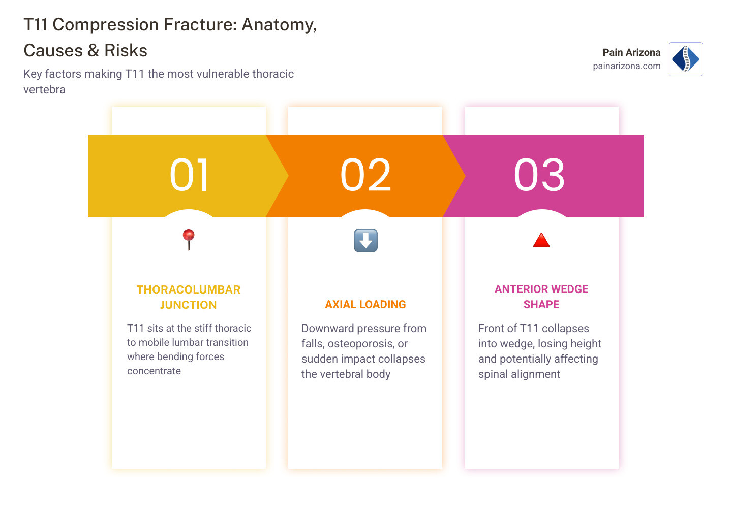
To understand why the T11 is so prone to breaking, we have to look at the “physics” of your back. The thoracic spine (T1 through T12) is connected to your rib cage, making it relatively stiff and stable. However, just below T12 is the lumbar spine (L1 through L5), which is incredibly mobile and flexible.
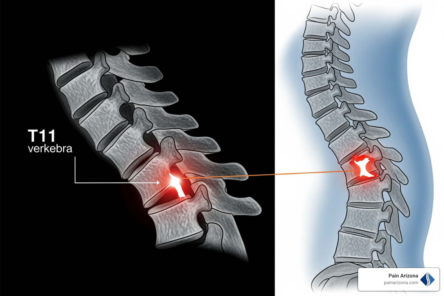
The T11 vertebra sits right at this “transition zone,” known as the thoracolumbar junction. When you bend, twist, or experience a sudden impact, the forces of that movement concentrate right where the stiff part of your back meets the flexible part. This is called axial loading. When the downward pressure exceeds the strength of the vertebral body, the front part of the bone collapses, creating an anterior wedge shape.
There are several back pain causes that can lead to this collapse, ranging from chronic bone thinning to sudden accidents.
The Role of Osteoporosis and Bone Density
In our Phoenix area clinics, the most common reason we see a compression fracture of T11 is osteoporosis. This condition causes a “silent” thinning of the bone, particularly in the trabecular bone (the spongy internal part of the vertebra).
For many postmenopausal women, hormonal changes lead to an imbalance where bone resorption (breaking down old bone) happens faster than the body can create new bone. This leaves the vertebrae brittle. In severe cases, the bone becomes so fragile that everyday activities — like reaching for a grocery bag or even a strong cough — can cause the T11 to buckle. We treat this as one of the primary conditions related to aging and spinal health.
Traumatic Injuries and Underlying Conditions
While osteoporosis is the leading cause for older adults, younger patients often sustain these fractures through high-energy trauma. This includes:
- Motor vehicle accidents: The sudden “forward-bending” force of a seatbelt during a crash can snap the anterior part of the T11.
- Falls from height: Landing on your feet or buttocks sends a shockwave straight up the spine.
- Sports injuries: High-impact contact sports can occasionally cause enough force to fracture healthy bone.
Beyond trauma, certain medical conditions can weaken the vertebral body from the inside out. Metastatic cancer (cancer that has spread from elsewhere) or multiple myeloma are known to “eat away” at the bone density. Additionally, long-term corticosteroid use for other health issues is a significant risk factor, as these medications can drastically reduce bone strength over time.
Recognizing Symptoms and Clinical Presentation
How do you know if that mid-back ache is a fracture or just a pulled muscle? The symptoms of a compression fracture of T11 can be surprisingly sneaky.
The most common sign is acute pain located right in the middle of the back. Unlike a muscle strain, which might feel better when you move around, fracture pain often feels like a deep, “boring” ache that worsens when you stand or walk and feels significantly better when you lie flat on your back.
Other clinical signs include:
- Midline tenderness: Pain when someone presses directly on the spine at the T11 level.
- Height loss: If you’ve noticed you’re an inch or two shorter than you were last year, it’s often due to multiple vertebrae collapsing.
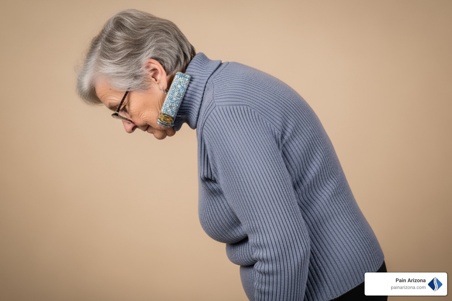
- Kyphosis: This is the medical term for a “hunchback” appearance. As the front of the T11 collapses into a wedge, it forces the spine to tilt forward.
For more on identifying these signs, check out our chronic back pain insights into persistent discomfort.
Identifying a Wedge Compression Fracture of T11
Most T11 fractures are “wedge” fractures. Imagine a cardboard box being stepped on only on one side — it squashes into a triangle. This anterior collapse means the back part of the vertebra remains at full height while the front part loses 20%, 30%, or even 50% of its height.
While this creates a loss of vertebral height, wedge fractures are generally considered “stable.” This means the bone isn’t in danger of sliding out of place or immediately damaging the spinal cord, though it still requires careful management to prevent the “wedge” from getting worse.
Neurological Red Flags and Complications
While rare in simple compression fractures, the bone can sometimes shatter or “burst.” If pieces of bone are pushed backward into the spinal canal, it can lead to nerve compression.
We call these “Red Flags,” and they require immediate medical attention:
- Numbness or tingling in the legs or “saddle area.”
- Sudden weakness or difficulty lifting your feet.
- Loss of bowel or bladder control.
These symptoms may indicate that the fracture has led to spinal stenosis, where the space for your nerves has become dangerously narrow.
Diagnostic Procedures and Severity Classification
When you visit us at Pain Arizona, we start with a thorough physical examination. We look at your posture, check your range of motion, and perform a neurological exam to ensure your nerves are firing correctly. However, imaging is the only way to confirm a compression fracture of T11.
- Spinal Radiographs (X-rays): Our first line of defense. X-rays are great for seeing the “wedge” shape and measuring how much height has been lost.
- MRI Imaging: This is the “gold standard” for acute fractures. An MRI shows “edema” (swelling) inside the bone. If the bone is swelling, we know the fracture is fresh. If there is no swelling, the fracture is old and likely already healed.
- CT Scan: We use this if we suspect a “burst” fracture, as it provides a 3D view of the bone fragments to see if they are close to the spinal cord.
- DEXA Scan: If the fracture happened without major trauma, we will likely order a bone density scan to check for underlying osteoporosis.
For a deeper dive into these tests, see our vertebral compression fracture guide.
Classification Systems for Spinal Stability
Doctors use specific “scorecards” to decide how to treat you.
- Genant Classification: Grades the fracture based on how much height is lost (Grade 1 is mild, Grade 3 is severe).
- TLICS Score: This looks at the “Thoracolumbar Injury Classification and Severity.” It helps us determine if the fracture is stable vs unstable.
- Burst Fracture vs Compression: A compression fracture only involves the front of the bone. A burst fracture involves the front and back, which is much more serious.
Treatment Options: From Conservative Care to Minimally Invasive Surgery
The good news is that many T11 fractures do not require surgery. Our goal at Pain Arizona is always to start with the least invasive option that will get you back to your life in Phoenix, Mesa, or Scottsdale.
| Treatment Type | Description | Best For |
|---|---|---|
| Conservative | Rest, NSAIDs, and bracing | Stable fractures with manageable pain |
| Minimally Invasive | Kyphoplasty or Vertebroplasty | Severe pain that doesn’t improve with rest |
| Surgical Fusion | Metal rods and screws | Unstable fractures or nerve damage |
Conservative Management
For many, the first 4 to 6 weeks involve “watchful waiting.” We use NSAIDs (like ibuprofen) or acetaminophen to manage the ache. We may also prescribe a TLSO brace (Thoracic-Lumbosacral Orthosis). This is a rigid brace that prevents you from bending forward, allowing the T11 bone to knit back together without being squashed further.
Minimally Invasive Surgery for Compression Fracture of T11
If the pain is debilitating or the bone continues to collapse, we may recommend vertebral augmentation. These are outpatient procedures performed through a tiny needle — no large incisions required!
- Vertebroplasty: We inject medical-grade bone cement directly into the fractured T11 to stabilize it.
- Kyphoplasty: First, we insert a small balloon into the T11 and inflate it to “lift” the collapsed bone back toward its original height. Then, we fill the space with bone cement.
Most patients feel a 50–90% reduction in pain within 24 to 48 hours of these procedures.
Physical Therapy and Rehabilitation
Whether you have surgery or heal naturally, rehab is vital. We focus on:
- Spinal extensors: Strengthening the muscles that keep you standing tall.
- Core strengthening: Building a “natural brace” around your spine.
- Posture training: Learning how to move without putting extra stress on the T11.
You can learn more about the minimally invasive spine surgery recovery time and what to expect during your journey.
Recovery Timeline and Long-Term Outlook
Healing a bone takes time. Generally, the 12-week healing mark is when the bone is considered “solid.”
- Weeks 1–4: Focus on pain control and activity modification. No heavy lifting (nothing heavier than a gallon of milk), no deep bending, and no twisting.
- Weeks 4–8: Gradual introduction of physical therapy and walking.
- Weeks 8–12: Weaning off the brace and increasing daily activities.
Long-term, we must address the root cause. If osteoporosis caused the fracture, we will work with you on bone health strategies, including calcium and Vitamin D supplementation, and possibly bone-building medications like bisphosphonates.
Frequently Asked Questions about T11 Fractures
Can a T11 compression fracture heal on its own?
Yes, many do! With a 3-month timeline, proper bracing, and activity restriction, the body can “scab over” the fracture with new bone. However, it will likely heal in its new, shorter “wedge” shape unless a kyphoplasty is performed to restore height.
When should I seek emergency medical attention?
You should head to the nearest Phoenix-area emergency room if you experience:
- Sudden, profound weakness in the legs.
- Loss of sensation in the “saddle” area or legs.
- High fever accompanied by back pain (which could indicate an infection).
- Sudden loss of bowel or bladder control.
What are the long-term effects of an untreated fracture?
An untreated compression fracture of T11 can lead to chronic pain and a permanent kyphosis (hunch). Severe hunching can eventually compress your internal organs, leading to respiratory issues (difficulty breathing) or digestive problems. Furthermore, having one fracture increases your risk of future fractures by five times.
Conclusion
A compression fracture of T11 can feel like a life-altering event, but with the right care, it doesn’t have to be. Whether you are in Glendale, Gilbert, or downtown Phoenix, our team at Pain Arizona is dedicated to providing evidence-based, compassionate care.
I’m Dr. Nasser, and we are here to help you navigate your recovery — from the first X-ray to your final physical therapy session. We offer the latest minimally invasive treatments to address the root cause of your pain and get you back to the activities you love.
If you suspect you have a spinal fracture or are struggling with chronic mid-back pain, don’t wait for the condition to worsen. We accept most insurance plans — please check with our office manager to see if your specific plan is accepted.
Ready to take the first step toward relief? View our Vertebral Compression Fracture Guide or contact one of our Greater Phoenix locations today to schedule your consultation.